Cusabio Ovis aries Recombinant

Introduction

The rumen microbiota plays an important role in the functional attributes of animals. These microbes are indispensable for the normal physiological development of the rumen and can also convert the vegetable polysaccharides in the grass into available milk and meat, making them very valuable to humans. Exploring the microbial composition and metabolites of the rumen throughout developmental stages is important for understanding ruminant nutrition and metabolism.

However, relatively few reports have investigated the microbiome and metabolites at developmental stages in ruminants. Using 16S rRNA gene sequencing, metabolomics, and high-performance liquid chromatography techniques, we compared rumen microbiota, metabolites, and short-chain fatty acids (SCFAs) between subadult Tibetan lambs and Ovis aries (sheep) Recombinant from Qinghai-Tibetan. Plateau. Bacteroidetes and Spirochaetae were more abundant in subadult sheep, while Firmicutes and Tenericutes were more abundant in young individuals. Subadult individuals had higher alpha diversity values ​​than young sheep.

Metabolomic analysis showed that essential amino acid content and functional pathways of related genes in the rumen were different between lambs and the subadult population. L-Leucine, which participates in the biosynthesis of valine, leucine, and isoleucine, was more abundant in lambs, while phenylethylamine, which participates in phenylalanine metabolism, was richer in subadults. Both rumen microbial community structures and metabolite profiles were affected by age, but rumen SCFA concentration was relatively stable between different age stages.
Some specific microbes (eg, Clostridium and Ruminococcaceae) were positively associated with L-leucine but negatively correlated with phenylethylamine, implying that rumen microbes may play different roles in metabolite production at different ages. Mantel test analysis showed that rumen microbiota was significantly correlated with metabolomics and SCFA profiles. Our results indicate the close relationship between microbial composition and metabolites, and also reveal different nutritional requirements for different ages in ruminants, which is of important importance for regulating animal nutrition and metabolism through the intervention of the microbiome.

Materials and methods

  • Animal feeding and sampling

Rumen contents were collected between February and July from the Plateau Modern Ecological Animal Husbandry Demonstration Area (100.9518°E, 36.9181°N) of Haibei. After anaesthetizing the sheep with diethyl ether and dissecting them, we obtained a total of 12 rumen samples from 1-month-old Tibetan sheep (the lamb group, abbreviated as ES; N = 6) and 6 months of age (the subadult group). ). , abbreviated as SS; N = 6) in the same cohort. Three servings (~5 g per serving) were taken from the contents of the fore, mid, and hind rumen and mixed well before sample collection.

These Tibetan sheep were all male. Subadult individuals grazed on pastures (main grass included Kobresia humilis, Oxytropis ochrocephala, Poa sp.) in the Qinghai-Tibet Plateau and fed commercial feed No. 8876 (Yongxing Ecological Agriculture and Animal Husbandry Development Co., Ltd. in Mengyuan County) at dusk. The main nutritional components of this commercial feed include crude protein ≥ 16%, crude fat ≥ 3%, crude fibre ≤ 8.0%, and crude ash ≤ 9.0%. The daily food consumption of the subadult individuals was 4.75 ± 0.32 kg.

The lambs were fed mainly milk, and also ate a small amount of grass (~0.72 ± 0.0.07 kg) and commercial feed (~0.54 ± 0.0.12 kg) mentioned above. Drinking water was freely available to these Tibetan sheep. The bodyweight of the lamb and subadult groups was 15.75 ± 5.90 and 26.35 ± 4.01 kg, respectively. After harvest, the ruminal content was immediately divided into three parts on ice for the subsequent microbiome, metabolome, and short-chain fatty acid (SCFA) analysis, and temporarily kept in a portable refrigerator at -20 °C in the field. . Finally, all samples were transferred to our laboratory within 24 h and stored in a -40 °C refrigerator.

  • Metabolomics measurement

100 mg of the ruminal contents were transferred to 5 ml centrifuge tubes and then 500 μL of ddH2O (4 °C) was added to the tubes. The mixture was thoroughly mixed by vortexing for 60 sec. Thereafter, 1000 ul of methanol (precooled to -20 °C) was added to the samples, and the mixed liquids were stirred for 30 s. Thereafter, we placed the tubes in an ultrasound machine at room temperature for 10 minutes and then stewed them for 30 minutes on ice. Samples were centrifuged for 10 min at 14,000 rpm at 4°C, and then 1.2 mL of supernatant was transferred to a new centrifuge tube.
The samples were blow-dried by concentration in vacuo. The samples were then dissolved using 400 μl of aqueous methanol solution (1:1, 4 °C) and subjected to 0.22 μm membrane filtration. For quality control (QC) samples, 20 μL of prepared samples were drawn and pooled. These quality control samples were used to control deviations in the analytical results of these pool mixes. Finally, the samples were ready for LC-MS detection (Waters, Milford, MA, USA). More detailed methods for LC-MS procedures are described in the previous report.

The original data obtained were converted to mzXML format using the Proteowizard software (v3.0.8789). The metabolomics data was then subjected to peak identification, filtering and alignment using the XCMS package in R (v3.3.2). In order to compare data of different magnitude, the peak area was normalized for further statistical analysis.

  • Measurement of short-chain fatty acids

Short-chain fatty acid (SCFA) profiles of ruminal content samples were measured using an Agilent 1100 series high-performance liquid chromatography (HPLC) system (Agilent Technologies, Santa Clara, CA, USA). We measured the concentration of acetate, propionate, butyrate, isobutyrate, valerate, isovalerate, and hexanoate using an Alltech IOA-2000 organic acid column. Detailed procedures for measuring SCFA.

Conclusion

In conclusion, we found that both rumen microbial community structures and metabolite profiles of Tibetan sheep were distinct at different ages, but rumen SCFA concentration was relatively stable between the two age stages. In particular, the metabolomic analysis showed that rumen essential amino acid nutritional requirements were different between lambs and subadult Tibetan ewes.

We found that L-Leucine, which is involved in the biosynthesis of valine, leucine, and isoleucine, was more abundant in lambs, while phenylethylamine, which is involved in phenylalanine metabolism, was richer in subadults. Furthermore, rumen microbiota was associated with metabolomics and SCFAs, indicating the close relationship between microbial composition and metabolites. These results have important significance for regulating animal nutrition and health through the intervention of the microbiome.

Cusabio N-terminal 10xHis-GST-tagged Recombinant

Abstract

Key assays in enzymology for biochemical characterization of proteins in vitro require high concentrations of the purified protein of interest. Protein purification protocols must combine efficiency, simplicity, and cost-effectiveness. Here, we describe the GST-His method as a new small-scale affinity purification system for recombinant proteins, based on an N-terminal glutathione sepharose (GST) tag and a C-terminal 10xHis tag, both fused to the protein of GST. interest. The latter construct is used to generate baculoviruses, for infection of Sf9-infected cells for protein expression.

GST is a fairly long tag (29 kDa) that serves to ensure the efficiency of the purification. However, it could influence the physiological properties of the protein. Therefore, it is subsequently cleaved from the protein using the PreScission enzyme. To ensure maximum purity and remove cleaved N-terminal 10xHis-GST-tagged Recombinant, we added a second affinity purification step based on the comparatively small His-Tag. Importantly, our technique relies on two different tags flanking the two ends of the protein, which is an efficient tool to remove degraded proteins and thus enriches full-length proteins.

The method presented here does not require an expensive instrument setup, such as FPLC. In addition, we incorporated MgCl2 and ATP washes to remove heat shock protein impurities and nuclease treatment to remove contaminating nucleic acids. In summary, the combination of two different flanking tags at the N- and C-terminus and the ability to cleave one of the tags ensures the recovery of a full-length, highly purified protein of interest.

Keywords: glutathione S-transferase (GST), pGEX, protein expression, protein purification, thrombin, factor Xa, fusion tags

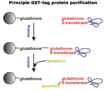
Affinity purification of GST fusion protein

Soluble GST fusion proteins are easily purified using an immobilized glutathione sepharose column. There are several immobilized glutathione chromatography media options available to purify soluble GST fusion proteins from bacterial cell lysates. The protocol described below is adapted from the manufacturer’s recommendation using Glutathione Sepharose 4B poured onto a column and using a peristaltic pump to control flow rates. Protease inhibitors and reducing agents should be added to the buffers as required to minimize proteolysis of the fusion protein. One exception is that serine protease inhibitors must be removed from the glutathione buffer prior to enzymatic removal of the GST moiety, as they will inhibit enzyme activity.

Save a small aliquot from each purification step for SDS-PAGE analysis to monitor fusion protein location throughout the purification. A given column or batch of resin should be used exclusively with a single fusion protein to minimize potential cross-contamination. As an alternative to column purification, a protocol describing batch purifications. Batch purifications are quick and easy, but often the yield and purity of the protein obtained will be somewhat less than that obtained by chromatographic separations. To minimize proteolysis, all protein purification steps should be carried out at 4°C, unless otherwise indicated.

  • Resuspend and pour 20 mL of Glutathione Sepharose 4B resin into a 2.5 × 8 cm column.
  • Thoroughly wash Glutathione Sepharose with 5–10 bed volume PBS at 1.5 mL/min to remove ethanol stock solution.
  • Resuspend pelleted E. coli cells in 15 ml cold Lysis buffer (cells can be fresh or thawed frozen cell pellets).
  • Lyse cells by sonication on ice (~10 times for 10 seconds each with 1-minute rest between bursts to minimize heating of the sample). Save 50 μl of lysate for on-gel analysis and transfer the rest to a 60 ml centrifuge tube.
  • Centrifuge the lysate at 48,000 × g for 20 min at 4 °C.
  • Decant the supernatant into a clean 50 mL centrifuge tube.
  • Resuspend the pellet in 15 ml PBS buffer using a Dounce homogenizer.
  • Run 5-10 μl of the lysate, supernatant, and resuspended pellet on an SDS-PAGE gel to verify that the fusion protein is in the supernatant fraction. If the fusion protein is in the pellet fraction, for tips on improving soluble protein expression for methods of extracting protein from inclusion bodies.
  • Load the soluble fusion protein onto the equilibrated glutathione sepharose column using a flow rate of 0.1 mL/min. Collect fractions and run gels to verify fusion protein binds to column and capacity has not been exceeded. If the fusion protein binds poorly to the resin, for various possible remedies.
  • Wash the column with 5–10 bed volumes of PBS/EDTA/PMSF using a flow rate of 1.5 mL/min.
  • Wash the column with 10-bed volumes of PBS/EDTA using a flow rate of 1.5 mL/min.
  • Elute the fusion protein with glutathione buffer using a flow rate of 0.3 mL/min. Fractions can be monitored using A280 and SDS-PAGE analysis. Pool fractions containing the GST fusion protein. The protein can be stored at 4°C and should be ~90% pure at this stage. If problems are found eluting the fusion protein. If high levels of contamination are present, for troubleshooting tips.

Enzymatic cleavage to remove the GST affinity tag

Depending on the vector chosen, the GST affinity tag can be removed with thrombin, factor Xa, or PreScission protease, either in solution or while still bound to the column matrix. Solution cleavage offers the advantage of greater control over optimizing cleavage conditions, such as temperature, enzyme-to-substrate ratio, incubation duration, and buffer conditions. An advantage of column cleavage is the high level of purity obtained, but this comes at the expense of generally low yield due to less efficient cleavage by proteases and less control of digestion conditions.

Digestion can be performed in the glutathione buffer used to elute the protein from the affinity matrix as long as there are no serine protease inhibitors in this buffer. After incubation, the enzyme can be inhibited using a variety of protease inhibitors or removed using a HiTrap Benzamidine column. Separation of the target protein and the GST moiety can be achieved by further chromatography on the glutathione Sepharose column (after dialysis in PBS buffer) to remove GST and any uncleaved fusion protein. For information on the digestion of GST fusion proteins while bound to column matrix (use with PreScission Protease is recommended).

  • Add the appropriate amount of thrombin or factor Xa to the affinity purified fusion protein and incubate at 37°C (thrombin) or 25°C (factor Xa) for the desired time.
  • Inactivate the enzyme by adding 0.3 mM PMSF (final concentration) to the sample. To ensure complete inhibition, incubate the sample for 15 min at 37°C for thrombin or 30 min at 25°C for factor Xa.
  • Dialyze the sample against PBS/EDTA/PMSF twice using 2 L per dialysis for a minimum of 4 h for each dialysis.
  • Centrifuge the dialyzed sample for 20 min at 4,000 × g to remove any precipitated material that may have formed during the digestion or dialysis steps. At this point, the sample can be reapplied to the glutathione Sepharose column to remove the remaining GST and any undigested fusion proteins.

.

Cusabio Apoptosis Recombinants

Abstract

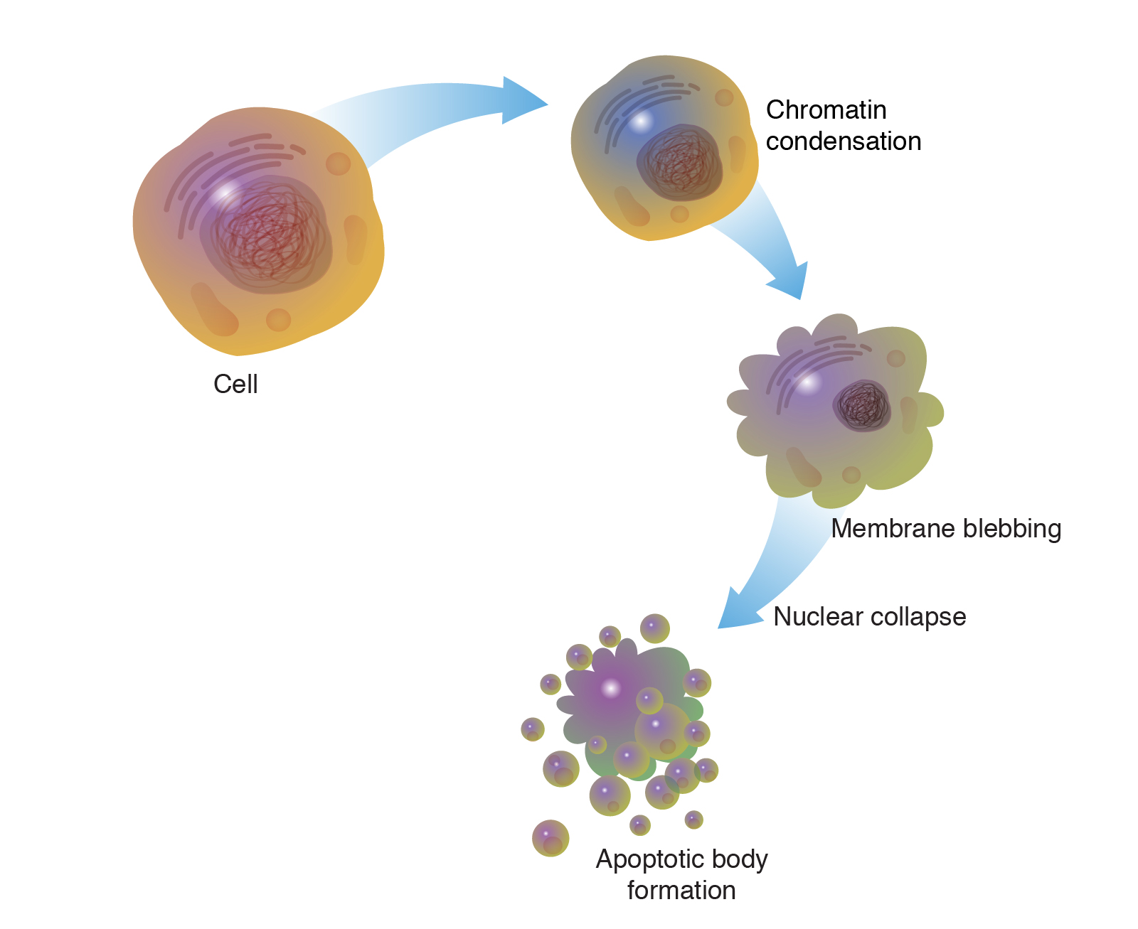
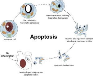
The process of programmed cell death, or apoptosis, is generally characterized by distinct morphological features and energy-dependent biochemical mechanisms. Apoptosis Recombinants is considered a vital component of several processes including normal cell turnover, proper development and function of the immune system, hormone-dependent atrophy, embryonic development, and chemical-induced cell death. Inappropriate apoptosis (either too little or too much) is a factor in many human conditions, including neurodegenerative diseases, ischemic damage, autoimmune disorders, and many types of cancer. The ability to modulate the life or death of a cell is recognized for its immense therapeutic potential.

Therefore, research continues to focus on the elucidation and analysis of the cell cycle machinery and signalling pathways that control cell cycle arrest and apoptosis. To that end, the field of apoptosis research has advanced at an alarmingly fast rate. Although many of the key apoptotic proteins have been identified, the molecular mechanisms of action or inaction of these proteins remain to be elucidated. The objective of this review is to provide an overview of current knowledge about the apoptosis process, including morphology, biochemistry, the role of apoptosis in health and disease, detection methods, as well as a discussion of possible ways apoptotic alternatives.

Keywords: Apoptosis, programmed cell death, intrinsic/extrinsic pathway, granzyme A/B, perforin, autophagy

Distinguish apoptosis from necrosis

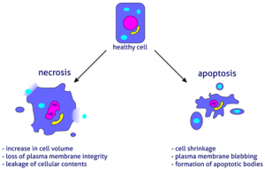
The alternative to apoptotic cell death is necrosis, which is considered a toxic process in which the cell is a passive victim and follows an energy-independent mode of death. But since necrosis refers to the degradative processes that occur after cell death, it is considered by some to be an inappropriate term to describe a mechanism of cell death. Therefore, oncosis is used to describe a process that leads to necrosis with karyolysis and cell swelling, while apoptosis leads to cell death with cell shrinkage, pyknosis, and karyorrhexis.

Therefore, the terms “oncotic cell death” and “oncotic necrosis” have been proposed as alternatives to describe cell death that is accompanied by cellular inflammation, but these terms are not widely used at this time (Majno and Joris, 1995; Levin et al., 1999). Although the mechanisms and morphologies of apoptosis and necrosis differ, these two processes overlap. Evidence indicates that necrosis and apoptosis represent morphological expressions of a shared biochemical network described as the “apoptosis-necrosis continuum” (Zeiss, 2003).

For example, two factors that will convert an ongoing apoptotic process into a necrotic process include a decrease in the availability of caspases and intracellular ATP (Leist et al., 1997; Denecker et al., 2001). Whether a cell dies by necrosis or apoptosis depends in part on the nature of the cell death signal, the type of tissue, the stage of tissue development, and the physiological milieu (Fiers et al., 1999; Zeiss, 2003).

Using conventional histology, it is not always easy to distinguish apoptosis from necrosis, and they can occur simultaneously depending on factors such as the intensity and duration of the stimulus, the degree of ATP depletion, and the availability of caspases (Zeiss, 2003). Necrosis is a passive and uncontrolled process that usually affects large fields of cells, while apoptosis is controlled and energy-dependent and can affect individual cells or groups of cells. Necrotic cell injury is mediated by two main mechanisms; interference with the cell’s energy supply and direct damage to cell membranes.

Physiological apoptosis

The role of apoptosis in normal physiology is as significant as that of its counterpart, mitosis. It demonstrates a complementary but opposite role to mitosis and cell proliferation in the regulation of various cell populations. It is estimated that to maintain homeostasis in the adult human body, about 10 billion cells are produced each day just to balance those that die by apoptosis (Renehan et al., 2001). And that number can increase significantly when apoptosis increases during normal development and ageing or during disease.

Apoptosis is critically important during various developmental processes. For example, both the nervous system and the immune system arise through the overproduction of cells. This initial overproduction is followed by the death of those cells that fail to establish functional synaptic connections or productive antigen specificities, respectively (Nijhawan et al., 2000; Opferman and Korsmeyer, 2003).

Apoptosis is also necessary to rid the body of cells invaded by pathogens and is a vital component of wound healing, as it is involved in the removal of inflammatory cells and the evolution of granulation tissue into scar tissue (Greenhalgh, 1998). . Dysregulation of apoptosis during wound healing can lead to pathological forms of scarring, such as excessive scarring and fibrosis. Apoptosis is also required to eliminate activated or autoaggressive immune cells either during maturation in central lymphoid organs (bone marrow and thymus) or in peripheral tissues (Osborne, 1996).

In addition, apoptosis is critical for remodellings in the adult, such as follicular atresia of the postovulatory follicle and postweaning mammary gland involution, to name a few examples (Tilly, 1991; Lund et al., 1996). Also, as organisms age, some cells begin to deteriorate at a faster rate and are eliminated by apoptosis. One theory is that oxidative stress plays a major role in the pathophysiology of age-induced apoptosis through accumulated free radical damage to mitochondrial DNA (Harman, 1992; Ozawa, 1995). It is clear that apoptosis has to be tightly regulated, as too little or too much cell death can lead to pathologies, including developmental defects, autoimmune diseases, neurodegeneration or cancer.

Apoptosis inhibition

There are many pathological conditions that exhibit excessive apoptosis (neurodegenerative diseases, AIDS, ischemia, etc.) and can therefore benefit from artificial inhibition of apoptosis. As our understanding of the field evolves, the identification and exploitation of new targets remain a considerable focus of attention (Nicholson, 2000). A shortlist of possible methods of anti-apoptotic therapy includes stimulation of the IAP (inhibitor of apoptosis) protein family, caspase inhibition, PARP (poly [ADP-ribose] polymerase) inhibition, stimulation of the PKB/Akt (protein kinase B) pathway and inhibition of Bcl-2 proteins.

The IAP family of proteins is perhaps the most important regulator of apoptosis due to the fact that it regulates both the intrinsic and extrinsic pathways (Deveraux and Reed, 1999). Eight human IAP proteins have now been identified, although XIAP (X-linked mammalian apoptosis inhibitory protein) and survivin remain the best-known members (Silke et al., 2002; Colnaghi et al., 2006). Until now, members of the IAP family have been investigated as therapeutic targets for the treatment of stroke, spinal cord injury, multiple sclerosis, and cancer.

The synthetic non-specific caspase inhibitor z-VAD-fmk was shown to reduce the severity of myocardial reperfusion injury in rat and mouse models of myocardial infarction (Mocanu et al., 2000). Specific inhibitors of caspase activity may also be of benefit. ICE (interleukin-1 beta converting enzyme), also called caspase I, is a cysteine ​​protease that appears to mediate intracellular protein degradation during apoptosis (Livingston, 1997). ACE inhibitors have been developed to treat rheumatoid arthritis and other inflammatory conditions by reducing interleukin 1β (Le and Abbenante, 2005).

Due to the dual role of PARP-1 in both DNA repair and apoptosis, pharmacological use of PARP-1 inhibitors may attenuate ischemic and inflammatory cell and organ injury or may increase the cytotoxicity of antitumor agents ( Graziani and Szabo, 2005). Recent research with PARP-1 knockout mice indicates that the use of PARP-1 inhibitors may be an effective therapy for injury associated with myocardial ischemia and reperfusion injury (Zhou et al., 2006). Infusion of insulin-like growth factor 1 (IGF-1), which stimulates PKB/Akt signalling and promotes cell survival, was shown to be beneficial in animal models of myocardial ischemia (Fujio et al., 2000).

Other studies with transgenic models of cardiac ischemia and global cerebral ischemia indicate that inhibition of Bax expression and/or function can prevent cytochrome c release from mitochondria, inhibit mitochondrial membrane potential lowering, and protect cells against apoptosis (Hochhauser et al., 2003; Hetz et al., 2005). The potential therapeutic modalities mentioned here represent just a few of the past and current research efforts in this field. As the molecular and biochemical complexities of apoptosis are elucidated, new therapeutic strategies will continue to evolve.

Conclusions

Apoptosis is considered a carefully regulated energy-dependent process, characterized by specific morphological and biochemical features in which caspase activation plays a central role. Although many of the key apoptotic proteins that are activated or deactivated in apoptotic pathways have been identified, the molecular mechanisms of action or activation of these proteins are not fully understood and are the subject of continuing research.

The importance of understanding the mechanical machinery of apoptosis is vital because programmed cell death is a component of both health and disease, being initiated by various physiological and pathological stimuli. Furthermore, the widespread involvement of apoptosis in the pathophysiology of disease lends itself to therapeutic intervention at many different checkpoints. Understanding the mechanisms of apoptosis and other variants of programmed cell death at the molecular level provides deeper insights into various disease processes and thus may influence therapeutic strategy.