Imaging stroboscopico ultraveloce con strisce RF in un microscopio elettronico a trasmissione commerciale
    1. Lo sviluppo della microscopia elettronica ultraveloce (UEM), in particolare l’imaging stroboscopico, ha portato lo studio della dinamica strutturale a un nuovo livello, superando i limiti spaziali della spettroscopia ultraveloce e i limiti di tempo della tradizionale TEM. La combinazione dei concetti che governano entrambe le tecniche ha consentito la visualizzazione diretta della dinamica con risoluzioni spazio-temporali nel regime del picosecondo-nanometro. Qui spostiamo i limiti  dell’imaging del fascio di elettroni pulsati tramite la deflessione trasversale indotta da RF basata su un generatore di impulsi a striscia di frequenza regolabile da 200 keV di nuova concezione.
    2. Presentiamo una risoluzione spaziale di 0,2 nm e spieghiamo le mappe di induzione dello spin magnetico mediante microscopia di fase. Presentiamo anche misurazioni della coerenza del fascio ed estendiamo il nostro studio utilizzando le modalità di respirazione del pettine di silicio di eccitazione RF, che forniscono una migliore tempistica tra il treno di impulsi di elettroni e il campo elettrico. È stato inoltre sviluppato un nuovo dispositivo RF con impedenza corrispondente al segnale RF per ridurre al minimo la perdita di potenza di trasmissione ai campioni e le sue prestazioni sono paragonate a quelle di un dispositivo campione convenzionale.
    Test del lettore di schede fototermico utilizzando una  striscia reattiva in oro colloidale disponibile in commercio per il rilevamento quantitativo rapido dei rischi alimentari

    • La semplice e veloce striscia reattiva in oro colloidale disponibile in commercio può essere utilizzata solo per il rilevamento qualitativo o semiquantitativo con scarsa rilevabilità e risultati falsi negativi. Questo documento sviluppa una striscia reattiva fototermica che combina la striscia reattiva con un lettore di schede fototermico portatile per ottenere un rilevamento quantitativo con un’eccellente rilevabilità. Secondo l’effetto fototermico prodotto dalle nanoparticelle d’oro (GNP) catturate sulla linea di test, il segnale potrebbe essere catturato da un lettore.
    • Sono stati studiati tredici rischi alimentari, inclusi residui di farmaci veterinari e residui di pesticidi; la rilevabilità fototermica nei campioni effettivi era di circa 23 (paration metilico), 7 (enrofloxacina), 6 (sarafloxacina), 8 (sulfadiazina), 12 (sulfametazina), 7 (paraquat), 6 (verde malachite), 11 (amantadina), 13 (nitrofurazone), 6 (dietilstilbestrolo), 12 (estriolo), 21 (estrone) e 26 (17β-estradiolo) volte meglio del rilevamento visivo. I nostri risultati hanno mostrato che la striscia reattiva fototermica può essere utilizzata per il rilevamento sensibile, rapido e quantitativo dei residui di rischio alimentare.

    Acetato di cellulosa e nanocomposito di cellulosa immobilizzati con il cromoforo DCDHF-idrazone verso una   striscia reattiva intelligente  per il rilevamento colorimetrico del dietilclorofosfato come fattore nervoso mimico

    1. L’esposizione agli agenti nervini, che di solito sono gas incolori e inodori, può causare insufficienza d’organo, paralisi e persino morte rapida. Il dietilclorofosfato (DCP) è stato riconosciuto come uno dei più noti imitatori di fattori nervosi nel combattimento chimico. Nel presente studio, presentiamo una semplice strategia per sviluppare una  striscia microporosa portatile e reversibile basata su nanocompositi per il rilevamento a occhio nudo di DCP in pochi secondi. Il cromoforo di dicianometilene diidrofuranidrazone (DCDHF-H) è stato sintetizzato mediante una facile reazione di accoppiamento azoico e incapsulato   in situ  durante la preparazione di nanocompositi di acetato di cellulosa / nanowiskers di cellulosa / idrazone (CA-CNW-H).
    2. Questi nanocompositi CA-CNW-H hanno mostrato uno spostamento batocromico dell’intensità di assorbimento di circa 142 nm da 438 a 580 nm con l’aumento della concentrazione di DCP. L’attuale striscia sensore CA-CNW-H ha mostrato un limite di rilevamento per DCP compreso tra 25 e 200 ppm. Il cambiamento di colore di CA-CNW-H da giallo a viola dopo l’esposizione a DCP è stato rilevato dall’analisi di CIE Lab. Sono state esaminate la morfologia, la cristallinità fibrosa, la stabilità termica e le proprietà meccaniche delle strisce di sensori CA-CNW-H preparate.

    Sviluppo di una test card basata su strisce immunocromatografiche d’oro colloidale per la rilevazione rapida di anticorpi contro Theileria equi e Babesia caballi.

    1. La piroplasmosi equina (EP) è un problema serio nell’industria equina e il controllo dell’EP è fondamentale per il commercio internazionale di cavalli. L’EP è causato da due protozoi apicomplessi, Theileria equi e Babesia caballi. Metodi rapidi e accurati adatti per rilevare questi parassiti sul campo sono fondamentali per controllare l’infezione e la diffusione di EP. In questo studio abbiamo sviluppato una scheda per la rilevazione degli anticorpi contro T. equi e B. caballi basata su due strisce immunocromatografiche in oro colloidale secondo il principio del double sandwich antigen. Le proteine ​​dell’antigene 1 (EMA1) dell’equimerozoite e la proteina rhoptry BC48 sono comunemente usate come antigeni diagnostici rispettivamente contro T. equi e B. caballi. La proteina EMA1 o BC48 purificata marcata con oro colloidale è stata utilizzata come rivelatore sulla striscia,
    2. Il protocollo richiede da 10 a 15 minuti e non richiede apparecchiature o prodotti chimici specializzati e un test può rilevare due agenti patogeni EP su una scheda. I test di specificità hanno confermato la mancanza di reattività crociata con sieri positivi per i comuni patogeni equini. Utilizzando un kit commerciale per l’immunosorbente enzimatico (cELISA) per il confronto,  476 campioni clinici sono stati testati mediante card. I tassi di coincidenza erano rispettivamente del 96,43% e del 97,90% per T. equi e B. caballi. Il feedback delle prove sul campo è stato costantemente positivo, suggerendo che questo strumento diagnostico può essere utile per controllare la diffusione di T. equi e B. caballi.
    3. La piroplasmosi equina (EP), causata da Theileria equi e Babesia caballi, è una grave malattia trasmessa dalle zecche nei cavalli che si verifica nella maggior parte del mondo. Il PE è considerato una malattia soggetta a denuncia dall’Organizzazione mondiale per la salute animale (OIE). La diagnosi accurata e la differenziazione di T. equi e B. caballi sono molto importanti per la prevenzione, il controllo e il trattamento dell’EP. Ecco perché abbiamo sviluppato  il test immunocromatografico Colloidal Gold Sandwich (GICG) per la rilevazione di T. equi e B. caballi. Due strisce GICG sono state impilate una accanto all’altra su una scheda di rilevamento di T. equi e B. caballi e in un test è stato possibile rilevare due agenti patogeni EP. Il metodo era semplice, veloce e specifico per la rilevazione di EP; pertanto, rispetto ai metodi precedenti, questo metodo è più adatto per la diagnostica dei patogeni sul campo.

    Strisce  immunocromatografiche fluorescenti ad alta specificità nel tempo per la rilevazione simultanea di diversi pesticidi organofosforici in base alla strategia della sonda indiretta

    • L’uso indiscriminato di pesticidi organofosforici (PO) rappresenta un’enorme minaccia per la sicurezza alimentare, la salute umana e l’ambiente. Poiché esistono molti tipi di pesticidi, individuarli è una sfida. Il rilevamento simultaneo di più organofosfati in un test è attualmente un problema da risolvere. In questo articolo,  è stata preparata una striscia di immunocromatografia a fluorescenza a risoluzione temporale (TRFIA) utilizzando anticorpi OP ad ampia specificità (Abs) come elemento di riconoscimento. Gli addominali sono stati combinati con microsfere di lattice di ossido di europio con anticorpi anti-topo di pecora (SaMIgG) per creare una sonda indiretta.
    • Questa strategia può realizzare efficacemente l’amplificazione del segnale e può risparmiare la quantità e proteggere l’attività degli addominali. Al rilevamento, è stato osservato un cambiamento di colore della linea del test (linea T) per la valutazione qualitativa alla luce UV (365 nm). Le immagini campione positive sono state quindi analizzate utilizzando ImageJ per completare il rilevamento quantitativo. In condizioni di progettazione e funzionamento ottimali, il limite di rilevamento della striscia può raggiungere 0,53 ng g  -1  .

Protein A Coated - 96 well plates - 8 Well Breakable Strips on single well holding frame - Clear PS

MG06F-PA/W Biomat 1 plate 30.36 EUR

Protein G Coated - 96 well plates - 8 Well Breakable Strips on single well holding frame - Clear PS

MG09F-PG Biomat 10 plates(2X5 plates in a transparent bag) 22.22 EUR

Protein G Coated - 96 well plates - 8 Well Breakable Strips on single well holding frame - Clear PS

MG09F-PG/W Biomat 1 plate 30.36 EUR

Wheat Germ Coated - 96 well plates - 8 Well Breakable Strips on single well holding frame - Clear PS

MG11F-WG Biomat 10 plates(2X5 plates in a transparent bag) 21.01 EUR

Wheat Germ Coated - 96 well plates - 8 Well Breakable Strips on single well holding frame - Clear PS

MG11F-WG/W Biomat 1 plate 28.05 EUR

Neutravidin Coated - 96 well plates - 8 Well Breakable Strips on single well holding frame - Clear PS

MG21F-NA Biomat 10 plates(2X5 plates in a transparent bag) 22.22 EUR

Neutravidin Coated - 96 well plates - 8 Well Breakable Strips on single well holding frame - Clear PS

MG21F-NA/W Biomat 1 plate 26.84 EUR

Streptavidin Coated - 96 well plates - 8 Well Breakable Strips on single well holding frame - Clear PS

MG0STF-SA5/200 Biomat 1 plate 23.32 EUR

Streptavidin Coated - 96 well plates - 8 Well Breakable Strips on single well holding frame - Clear PS

MG0STF-SA5/200/W Biomat 1 plate 26.84 EUR

Protein A/G Coated - 96 well plates - 8 Well Breakable Strips on single well holding frame - Clear PS

MG08F-PAG Biomat 10 plates(2X5 plates in a transparent bag) 37.4 EUR

Protein A/G Coated - 96 well plates - 8 Well Breakable Strips on single well holding frame - Clear PS

MG08F-PAG/W Biomat 1 plate 51.37 EUR

Concanavalin A Coated - 96 well plates - 8 Well Breakable Strips on single well holding frame - Clear PS

MG07F-CON_A Biomat 1 plate 15.18 EUR

Concanavalin A Coated - 96 well plates - 8 Well Breakable Strips on single well holding frame - Clear PS

MG07F-CON_A/W Biomat 1 plate 20.46 EUR

Poly-L-lysine Coated - 96 well plates - 8 Well Breakable Strips on single well holding frame - Clear PS

MG12F-LYS-L Biomat 10 plates(2X5 plates in a transparent bag) 15.4 EUR

Heparin Catcher HC3 Coated - 96 well plates - 8 Well Breakable Strips on single well holding frame - Clear PS

MG20F-HC3 Biomat 10 plates(2X5 plates in a transparent bag) 18.7 EUR

Heparin Catcher HC3 Coated - 96 well plates - 8 Well Breakable Strips on single well holding frame - Clear PS

MG20F-HC3/W Biomat 1 plate 21.78 EUR

Poly-L-arginine Coated - 96 well plates - 8 Well Breakable Strips on single well holding frame - Clear PS

MG13F-AR Biomat 10 plates(2X5 plates in a transparent bag) 21.01 EUR

Heparin Catcher HC1 Coated - 96 well plates - 8 Well Breakable Strips on single well holding frame - Clear PS

MG18F-HC1 Biomat 10 plates(2X5 plates in a transparent bag) 17.49 EUR

Heparin Catcher HC1 Coated - 96 well plates - 8 Well Breakable Strips on single well holding frame - Clear PS

MG18F-HC1/W Biomat 1 plate 21.56 EUR

Heparin Catcher HC2 Coated - 96 well plates - 8 Well Breakable Strips on single well holding frame - Clear PS

MG19F-HC2 Biomat 10 plates(2X5 plates in a transparent bag) 18.7 EUR

Heparin Catcher HC2 Coated - 96 well plates - 8 Well Breakable Strips on single well holding frame - Clear PS

MG19F-HC2/W Biomat 1 plate 21.78 EUR

Goat anti-Mouse IgG Coated - 96 well plates - 8 Well Breakable Strips on single well holding frame - Clear PS

MG25F-aM Biomat 10 plates(2X5 plates in a transparent bag) 32.67 EUR

Goat anti-Mouse IgG Coated - 96 well plates - 8 Well Breakable Strips on single well holding frame - Clear PS

MG25F-aM/W Biomat 1 plate 38.5 EUR

Goat anti-Rabbit IgG Coated - 96 well plates - 8 Well Breakable Strips on single well holding frame - Clear PS

MG26F-aR Biomat 10 plates(2X5 plates in a transparent bag) 28.05 EUR

Goat anti-Rabbit IgG Coated - 96 well plates - 8 Well Breakable Strips on single well holding frame - Clear PS

MG26F-aR/W Biomat 1 plate 33.88 EUR

Streptavidin High Binding Coated - 96 well plates - 8 Well Breakable Strips on single well holding frame - Clear PS

MGSTDF-SB75/100 Biomat 1 plate 25.74 EUR

Streptavidin High Binding Coated - 96 well plates - 8 Well Breakable Strips on single well holding frame - Clear PS

MGSTDF-SB75/100/W Biomat 1 plate 29.15 EUR

High Binding Capacity - 96 well plates - 12x8 Well Breakable Strips on single well holding frame (Clear)

BSV-MG01F-HB8 BioServUK 100 pcs 606 EUR

Medium Binding Capacity - 96 well plates - 12x8 Well Breakable Strips on single well holding frame (Clear)

BSV-MG0F-MB BioServUK 100 pcs 566 EUR

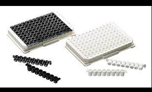
No Binding 96 well plates - 8 Well Breakable Strips on single well holding frame - Black PS

MGB03F2-NB Biomat 10 plates(2X5 plates in a transparent bag) 10.78 EUR

No Binding 96 well plates - 8 Well Breakable Strips on single well holding frame - White PS

MGW03F-NB Biomat 10 plates(2X5 plates in a transparent bag) 10.78 EUR

96 well plates - 12x8 Well Breakable Strips on single well holding frame

MGB0STF2-SA5-200 Biomat 10 plates(2X5 plates in a transparent bag) 265.07 EUR

96 well plates - 12x8 Well Breakable Strips on single well holding frame

MGB0STF2-SA5-200-W Biomat 10 plates(2X5 plates in a transparent bag) 299.09 EUR

96 well plates - 12x8 Well Breakable Strips on single well holding frame

MGB15F2-BIO-W Biomat 10 plates(2X5 plates in a transparent bag) 287.75 EUR

96 well plates - 12x8 Well Breakable Strips on single well holding frame

MGW0STF-SA5-200 Biomat 10 plates(2X5 plates in a transparent bag) 265.07 EUR

96 well plates - 12x8 Well Breakable Strips on single well holding frame

MGW0STF-SA5-200-W Biomat 10 plates(2X5 plates in a transparent bag) 299.09 EUR

96 well plates - 12x8 Well Breakable Strips on single well holding frame

MGW15F-BIO-W Biomat 10 plates(2X5 plates in a transparent bag) 287.75 EUR

96 well plates - 12x8 Well Breakable Strips on single well holding frame

MG0STF-SA5-200 Biomat 10 plates(2X5 plates in a transparent bag) 262.8 EUR

96 well plates - 12x8 Well Breakable Strips on single well holding frame

MG0STF-SA5-200-W Biomat 10 plates(2X5 plates in a transparent bag) 296.82 EUR

96 well plates - 12x8 Well Breakable Strips on single well holding frame

MG15F-BIO-W Biomat 10 plates(2X5 plates in a transparent bag) 285.48 EUR

High Binding 96 well plates - 8 Well Breakable Strips on single well holding frame - Black PS

MGB01F2-HB8 Biomat 1 plate 3.74 EUR

High Binding 96 well plates - 8 Well Breakable Strips on single well holding frame - White PS

MGW01F-HB8 Biomat 1 plate 3.74 EUR

Carboxylated - 96 well plates - 8 Well Breakable Strips on single well holding frame - Black PS

MGB04F2-COOH Biomat 10 plates(2X5 plates in a transparent bag) 14.3 EUR

Carboxylated - 96 well plates - 8 Well Breakable Strips on single well holding frame - White PS

MGW04F-COOH Biomat 10 plates(2X5 plates in a transparent bag) 14.3 EUR

Amine Treated - 96 well plates - 8 Well Breakable Strips on single well holding frame - Black PS

MGB02F2-AM1 Biomat 10 plates(2X5 plates in a transparent bag) 21.23 EUR

Medium Binding 96 well plates - 8 Well Breakable Strips on single well holding frame - Black PS

MGB0F2-MB Biomat 1 plate 3.52 EUR

Jacalin Coated - 96 well plates - 8 Well Breakable Strips on single well holding frame - Black PS

MGB10F2-JAC Biomat 10 plates(2X5 plates in a transparent bag) 17.82 EUR

Biotin Coated - 96 well plates - 8 Well Breakable Strips on single well holding frame - Black PS

MGB15F2-BIO Biomat 10 plates(2X5 plates in a transparent bag) 18.92 EUR

Biotin Coated - 96 well plates - 8 Well Breakable Strips on single well holding frame - Black PS

MGB15F2-BIO/W Biomat 1 plate 25.96 EUR

Amine Treated - 96 well plates - 8 Well Breakable Strips on single well holding frame - White PS

MGW02F-AM1 Biomat 10 plates(2X5 plates in a transparent bag) 21.23 EUR

Medium Binding 96 well plates - 8 Well Breakable Strips on single well holding frame - White PS

MGW0F-MB Biomat 1 plate 3.52 EUR

Jacalin Coated - 96 well plates - 8 Well Breakable Strips on single well holding frame - White PS

MGW10F-JAC Biomat 10 plates(2X5 plates in a transparent bag) 17.82 EUR

Biotin Coated - 96 well plates - 8 Well Breakable Strips on single well holding frame - White PS

MGW15F-BIO Biomat 10 plates(2X5 plates in a transparent bag) 18.92 EUR

Biotin Coated - 96 well plates - 8 Well Breakable Strips on single well holding frame - White PS

MGW15F-BIO/W Biomat 1 plate 25.96 EUR

Maleimide Coated - 96 well plates - 8 Well Breakable Strips on single well holding frame - Black PS

MGB22F2-MAL Biomat 10 plates(2X5 plates in a transparent bag) 28.27 EUR

Maleimide Coated - 96 well plates - 8 Well Breakable Strips on single well holding frame - White PS

MGW22F-MAL Biomat 10 plates(2X5 plates in a transparent bag) 28.27 EUR

Protein A Coated - 96 well plates - 8 Well Breakable Strips on single well holding frame - Black PS

MGB06F2-PA Biomat 10 plates(2X5 plates in a transparent bag) 22.44 EUR

Protein A Coated - 96 well plates - 8 Well Breakable Strips on single well holding frame - Black PS

MGB06F2-PA/W Biomat 1 plate 30.58 EUR

Protein G Coated - 96 well plates - 8 Well Breakable Strips on single well holding frame - Black PS

MGB09F2-PG Biomat 10 plates(2X5 plates in a transparent bag) 22.44 EUR

Protein G Coated - 96 well plates - 8 Well Breakable Strips on single well holding frame - Black PS

MGB09F2-PG/W Biomat 1 plate 30.58 EUR

Wheat Germ Coated - 96 well plates - 8 Well Breakable Strips on single well holding frame - Black PS

MGB11F2-WG Biomat 10 plates(2X5 plates in a transparent bag) 21.23 EUR

Wheat Germ Coated - 96 well plates - 8 Well Breakable Strips on single well holding frame - Black PS

MGB11F2-WG/W Biomat 1 plate 28.27 EUR

Protein A Coated - 96 well plates - 8 Well Breakable Strips on single well holding frame - White PS

MGW06F-PA Biomat 10 plates(2X5 plates in a transparent bag) 22.44 EUR

Protein A Coated - 96 well plates - 8 Well Breakable Strips on single well holding frame - White PS

MGW06F-PA/W Biomat 1 plate 30.58 EUR

Protein G Coated - 96 well plates - 8 Well Breakable Strips on single well holding frame - White PS

MGW09F-PG Biomat 10 plates(2X5 plates in a transparent bag) 22.44 EUR

Protein G Coated - 96 well plates - 8 Well Breakable Strips on single well holding frame - White PS

MGW09F-PG/W Biomat 1 plate 30.58 EUR

Wheat Germ Coated - 96 well plates - 8 Well Breakable Strips on single well holding frame - White PS

MGW11F-WG Biomat 10 plates(2X5 plates in a transparent bag) 21.23 EUR

Wheat Germ Coated - 96 well plates - 8 Well Breakable Strips on single well holding frame - White PS

MGW11F-WG/W Biomat 1 plate 28.27 EUR

Neutravidin Coated - 96 well plates - 8 Well Breakable Strips on single well holding frame - Black PS

MGB21F2-NA Biomat 10 plates(2X5 plates in a transparent bag) 22.44 EUR

Neutravidin Coated - 96 well plates - 8 Well Breakable Strips on single well holding frame - Black PS

MGB21F2-NA/W Biomat 1 plate 27.06 EUR

Neutravidin Coated - 96 well plates - 8 Well Breakable Strips on single well holding frame - White PS

MGW21F-NA Biomat 10 plates(2X5 plates in a transparent bag) 22.44 EUR

Neutravidin Coated - 96 well plates - 8 Well Breakable Strips on single well holding frame - White PS

MGW21F-NA/W Biomat 1 plate 27.06 EUR

Streptavidin Coated - 96 well plates - 8 Well Breakable Strips on single well holding frame - Black PS

MGB0STF2-SA5/200 Biomat 1 plate 23.65 EUR

Streptavidin Coated - 96 well plates - 8 Well Breakable Strips on single well holding frame - Black PS

MGB0STF2-SA5/200/W Biomat 1 plate 27.06 EUR

Streptavidin Coated - 96 well plates - 8 Well Breakable Strips on single well holding frame - White PS

MGW0STF-SA5/200 Biomat 1 plate 23.65 EUR

Streptavidin Coated - 96 well plates - 8 Well Breakable Strips on single well holding frame - White PS

MGW0STF-SA5/200/W Biomat 1 plate 27.06 EUR

Protein A/G Coated - 96 well plates - 8 Well Breakable Strips on single well holding frame - Black PS

MGB08F2-PAG Biomat 10 plates(2X5 plates in a transparent bag) 37.95 EUR

Protein A/G Coated - 96 well plates - 8 Well Breakable Strips on single well holding frame - Black PS

MGB08F2-PAG/W Biomat 1 plate 51.59 EUR

Protein A/G Coated - 96 well plates - 8 Well Breakable Strips on single well holding frame - White PS

MGW08F-PAG Biomat 10 plates(2X5 plates in a transparent bag) 37.95 EUR

Protein A/G Coated - 96 well plates - 8 Well Breakable Strips on single well holding frame - White PS

MGW08F-PAG/W Biomat 1 plate 51.59 EUR

Concanavalin A Coated - 96 well plates - 8 Well Breakable Strips on single well holding frame - Black PS

MGB07F2-CON_A Biomat 1 plate 15.4 EUR

Concanavalin A Coated - 96 well plates - 8 Well Breakable Strips on single well holding frame - Black PS

MGB07F2-CON_A/W Biomat 1 plate 20.79 EUR

Concanavalin A Coated - 96 well plates - 8 Well Breakable Strips on single well holding frame - White PS

MGW07F-CON_A Biomat 1 plate 15.4 EUR

Concanavalin A Coated - 96 well plates - 8 Well Breakable Strips on single well holding frame - White PS

MGW07F-CON_A/W Biomat 1 plate 20.79 EUR

Poly-L-lysine Coated - 96 well plates - 8 Well Breakable Strips on single well holding frame - Black PS

MGB12F2-LYS-L Biomat 10 plates(2X5 plates in a transparent bag) 15.73 EUR

Poly-L-lysine Coated - 96 well plates - 8 Well Breakable Strips on single well holding frame - White PS

MGW12F-LYS-L Biomat 10 plates(2X5 plates in a transparent bag) 15.73 EUR

Poly-L-arginine Coated - 96 well plates - 8 Well Breakable Strips on single well holding frame - Black PS

MGB13F2-AR Biomat 10 plates(2X5 plates in a transparent bag) 21.23 EUR

Heparin Catcher HC1 Coated - 96 well plates - 8 Well Breakable Strips on single well holding frame - Black PS

MGB18F2-HC1 Biomat 10 plates(2X5 plates in a transparent bag) 17.82 EUR

Heparin Catcher HC1 Coated - 96 well plates - 8 Well Breakable Strips on single well holding frame - Black PS

MGB18F2-HC1/W Biomat 1 plate 21.78 EUR

Heparin Catcher HC2 Coated - 96 well plates - 8 Well Breakable Strips on single well holding frame - Black PS

MGB19F2-HC2 Biomat 10 plates(2X5 plates in a transparent bag) 18.92 EUR

Heparin Catcher HC2 Coated - 96 well plates - 8 Well Breakable Strips on single well holding frame - Black PS

MGB19F2-HC2/W Biomat 1 plate 22 EUR

Heparin Catcher HC3 Coated - 96 well plates - 8 Well Breakable Strips on single well holding frame - Black PS

MGB20F2-HC3 Biomat 10 plates(2X5 plates in a transparent bag) 18.92 EUR

Heparin Catcher HC3 Coated - 96 well plates - 8 Well Breakable Strips on single well holding frame - Black PS

MGB20F2-HC3/W Biomat 1 plate 22 EUR

Poly-L-arginine Coated - 96 well plates - 8 Well Breakable Strips on single well holding frame - White PS

MGW13F-AR Biomat 10 plates(2X5 plates in a transparent bag) 21.23 EUR

Heparin Catcher HC1 Coated - 96 well plates - 8 Well Breakable Strips on single well holding frame - White PS

MGW18F-HC1 Biomat 10 plates(2X5 plates in a transparent bag) 17.82 EUR

Heparin Catcher HC1 Coated - 96 well plates - 8 Well Breakable Strips on single well holding frame - White PS

MGW18F-HC1/W Biomat 1 plate 21.78 EUR

Heparin Catcher HC2 Coated - 96 well plates - 8 Well Breakable Strips on single well holding frame - White PS

MGW19F-HC2 Biomat 10 plates(2X5 plates in a transparent bag) 18.92 EUR

Heparin Catcher HC2 Coated - 96 well plates - 8 Well Breakable Strips on single well holding frame - White PS

MGW19F-HC2/W Biomat 1 plate 22 EUR

Heparin Catcher HC3 Coated - 96 well plates - 8 Well Breakable Strips on single well holding frame - White PS

MGW20F-HC3 Biomat 10 plates(2X5 plates in a transparent bag) 18.92 EUR

Heparin Catcher HC3 Coated - 96 well plates - 8 Well Breakable Strips on single well holding frame - White PS

MGW20F-HC3/W Biomat 1 plate 22 EUR

Goat anti-Mouse IgG Coated - 96 well plates - 8 Well Breakable Strips on single well holding frame - Black PS

MGB25F2-aM Biomat 1 plate 32.89 EUR

Goat anti-Mouse IgG Coated - 96 well plates - 8 Well Breakable Strips on single well holding frame - Black PS

MGB25F2-aM/W Biomat 1 plate 38.72 EUR

Goat anti-Rabbit IgG Coated - 96 well plates - 8 Well Breakable Strips on single well holding frame - Black PS

MGB26F2-aR Biomat 1 plate 28.27 EUR

Goat anti-Rabbit IgG Coated - 96 well plates - 8 Well Breakable Strips on single well holding frame - Black PS

MGB26F2-aR/W Biomat 1 plate 34.1 EUR

Goat anti-Mouse IgG Coated - 96 well plates - 8 Well Breakable Strips on single well holding frame - White PS

MGW25F-aM Biomat 10 plates(2X5 plates in a transparent bag) 32.89 EUR

Goat anti-Mouse IgG Coated - 96 well plates - 8 Well Breakable Strips on single well holding frame - White PS

MGW25F-aM/W Biomat 1 plate 38.72 EUR

Goat anti-Rabbit IgG Coated - 96 well plates - 8 Well Breakable Strips on single well holding frame - White PS

MGW26F-aR Biomat 10 plates(2X5 plates in a transparent bag) 28.27 EUR

Goat anti-Rabbit IgG Coated - 96 well plates - 8 Well Breakable Strips on single well holding frame - White PS

MGW26F-aR/W Biomat 1 plate 34.1 EUR

Streptavidin High Binding Coated - 96 well plates - 8 Well Breakable Strips on single well holding frame - Black PS

MGBSTDF2-SB75/100 Biomat 1 plate 25.96 EUR

Streptavidin High Binding Coated - 96 well plates - 8 Well Breakable Strips on single well holding frame - Black PS

MGBSTDF2-SB75/100/W Biomat 1 plate 29.48 EUR

Streptavidin High Binding Coated - 96 well plates - 8 Well Breakable Strips on single well holding frame - White PS

MGWSTDF-SB75/100 Biomat 1 plate 25.96 EUR

Streptavidin High Binding Coated - 96 well plates - 8 Well Breakable Strips on single well holding frame - White PS

MGWSTDF-SB75/100/W Biomat 1 plate 29.48 EUR

High Binding Capacity - 96 well plates - 12x8 Well Breakable Strips on single well holding frame (Black)

BSV-MGB01F2-HB8 BioServUK 100 pcs 647 EUR

High Binding Capacity - 96 well plates - 12x8 Well Breakable Strips on single well holding frame (White)

BSV-MGW01F-HB8 BioServUK 100 pcs 647 EUR

Medium Binding Capacity - 96 well plates - 12x8 Well Breakable Strips on single well holding frame (Black)

BSV-MGB0F2-MB BioServUK 100 pcs 606 EUR

Medium Binding Capacity - 96 well plates - 12x8 Well Breakable Strips on single well holding frame (White)

BSV-MGW0F-MB BioServUK 100 pcs 606 EUR

Testosterone ELISA, Breakable Well

GWB-472DB9 GenWay Biotech 96 Tests Ask for price

8-well PCR Strip Tubes, Clear

27-425 Genesee Scientific Box of 125 Strips/Unit 67.53 EUR

Goat anti-Mouse, 1 x 8 Strip Well, Clear Coated 96 Well Plate, 1EA

X012-1EA Arbor Assays 1EA 48 EUR

Break-2-ways Plate Breakable - PK50

PCR1280 Scientific Laboratory Supplies PK50 454.95 EUR

No Binding 96 well plates - 8 Well Strips on 12 x 8 frame - Clear PS

MT03F4-NB Biomat 1 plate 10.56 EUR

Carboxylated - 96 well plates - 8 Well Strips on 12 x 8 frame - Clear PS

MT04F4-COOH Biomat 1 plate 14.08 EUR

High Binding 96 well plates - 8 Well Strips on 12 x 8 frame - Clear PS

MT01F4-HB8 Biomat 1 plate 3.52 EUR

Amine Treated - 96 well plates - 8 Well Strips on 12 x 8 frame - Clear PS

MT02F4-AM1 Biomat 1 plate 21.01 EUR

Biotin Coated - 96 well plates - 8 Well Strips on 12 x 8 frame - Clear PS

MT15F4-BIO Biomat 1 plate 18.7 EUR

Biotin Coated - 96 well plates - 8 Well Strips on 12 x 8 frame - Clear PS

MT15F4-BIO/W Biomat 1 plate 25.74 EUR

Medium Binding 96 well plates - 8 Well Strips on 12 x 8 frame - Clear PS

MT0F4-MB Biomat 1 plate 3.3 EUR

Jacalin Coated - 96 well plates - 8 Well Strips on 12 x 8 frame - Clear PS

MT10F4-JAC Biomat 1 plate 17.49 EUR

Maleimide Coated - 96 well plates - 8 Well Strips on 12 x 8 frame - Clear PS

MT22F4-MAL Biomat 1 plate 28.05 EUR

0.2ml 8-Strip Tubes & Flat Strip Caps, Clear

24-706 Genesee Scientific Box of 125 Tubes & Caps/Unit 74.53 EUR

Streptavidin Coated - 96 well plates - 8 Well Strips on 12 x 8 frame - Clear PS

MT0STF4-SA5/200 Biomat 1 plate 23.32 EUR

Streptavidin Coated - 96 well plates - 8 Well Strips on 12 x 8 frame - Clear PS

MT0STF4-SA5/200/W Biomat 1 plate 26.84 EUR

Neutravidin Coated - 96 well plates - 8 Well Strips on 12 x 8 frame - Clear PS

MT21F4-NA Biomat 1 plate 22.22 EUR

Neutravidin Coated - 96 well plates - 8 Well Strips on 12 x 8 frame - Clear PS

MT21F4-NA/W Biomat 1 plate 26.84 EUR

Protein A Coated - 96 well plates - 8 Well Strips on 12 x 8 frame - Clear PS

MT06F4-PA Biomat 1 plate 22.22 EUR

Protein A Coated - 96 well plates - 8 Well Strips on 12 x 8 frame - Clear PS

MT06F4-PA/W Biomat 1 plate 30.36 EUR

Protein G Coated - 96 well plates - 8 Well Strips on 12 x 8 frame - Clear PS

MT09F4-PG Biomat 1 plate 22.22 EUR

Protein G Coated - 96 well plates - 8 Well Strips on 12 x 8 frame - Clear PS

MT09F4-PG/W Biomat 1 plate 30.36 EUR

Wheat Germ Coated - 96 well plates - 8 Well Strips on 12 x 8 frame - Clear PS

MT11F4-WG Biomat 1 plate 21.01 EUR

Wheat Germ Coated - 96 well plates - 8 Well Strips on 12 x 8 frame - Clear PS

MT11F4-WG/W Biomat 1 plate 28.05 EUR

Protein A/G Coated - 96 well plates - 8 Well Strips on 12 x 8 frame - Clear PS

MT08F4-PAG Biomat 1 plate 37.4 EUR

Protein A/G Coated - 96 well plates - 8 Well Strips on 12 x 8 frame - Clear PS

MT08F4-PAG/W Biomat 1 plate 51.37 EUR

Concanavalin A Coated - 96 well plates - 8 Well Strips on 12 x 8 frame - Clear PS

MT07F4-CON_A Biomat 1 plate 15.18 EUR

Concanavalin A Coated - 96 well plates - 8 Well Strips on 12 x 8 frame - Clear PS

MT07F4-CON_A/W Biomat 1 plate 20.46 EUR

0.2mL Clear Polypropylene PCR Tubes 8 Well Strips - PK300

6542 Scientific Laboratory Supplies PK300 726.3 EUR

0.2ml 8-Strip Tubes and Dome Strip Caps, Clear

24-705 Genesee Scientific Box of 125 Tubes and Caps/Unit 69.27 EUR

Poly-L-lysine Coated - 96 well plates - 8 Well Strips on 12 x 8 frame - Clear PS

MT12F4-LYS-L Biomat 1 plate 15.4 EUR

Poly-L-arginine Coated - 96 well plates - 8 Well Strips on 12 x 8 frame - Clear PS

MT13F4-AR Biomat 1 plate 21.01 EUR

Heparin Catcher HC1 Coated - 96 well plates - 8 Well Strips on 12 x 8 frame - Clear PS

MT18F4-HC1 Biomat 1 plate 17.49 EUR

Heparin Catcher HC1 Coated - 96 well plates - 8 Well Strips on 12 x 8 frame - Clear PS

MT18F4-HC1/W Biomat 1 plate 21.56 EUR

Heparin Catcher HC2 Coated - 96 well plates - 8 Well Strips on 12 x 8 frame - Clear PS

MT19F4-HC2 Biomat 1 plate 18.7 EUR

Heparin Catcher HC2 Coated - 96 well plates - 8 Well Strips on 12 x 8 frame - Clear PS

MT19F4-HC2/W Biomat 1 plate 21.78 EUR

Heparin Catcher HC3 Coated - 96 well plates - 8 Well Strips on 12 x 8 frame - Clear PS

MT20F4-HC3 Biomat 1 plate 18.7 EUR

Heparin Catcher HC3 Coated - 96 well plates - 8 Well Strips on 12 x 8 frame - Clear PS

MT20F4-HC3/W Biomat 1 plate 21.78 EUR

Goat anti-Mouse IgG Coated - 96 well plates - 8 Well Strips on 12 x 8 frame - Clear PS

MT25F4-aM Biomat 1 plate 32.67 EUR

Goat anti-Mouse IgG Coated - 96 well plates - 8 Well Strips on 12 x 8 frame - Clear PS

MT25F4-aM/W Biomat 1 plate 38.5 EUR

Nickel Coated Plates, Clear, 12×8-Well Strips, White Frame

SP-16 ACROBIOSYSTEMS 1plate 310.3 EUR

Goat anti-Rabbit IgG Coated - 96 well plates - 8 Well Strips on 12 x 8 frame - Clear PS

MT26F4-aR/W Biomat 1 plate 33.88 EUR

Goat anti-Rabbit IgG Coated - 96 well plates - 8 Well Strips on 12 x 8 frame - Clear PS

MT26F4-aR_ Biomat 1 plate 28.05 EUR

Streptavidin High Binding Coated - 96 well plates - 8 Well Strips on 12 x 8 frame - Clear PS

MTSTDF4-SB75/100 Biomat 1 plate 25.74 EUR

Streptavidin High Binding Coated - 96 well plates - 8 Well Strips on 12 x 8 frame - Clear PS

MTSTDF4-SB75/100/W Biomat 1 plate 29.15 EUR

Streptavidin Coated Plates, Clear, 12×8-Well Strips, White Frame

SP-11 ACROBIOSYSTEMS 1plate 149.8 EUR

0.2ML THIN WALL CLEAR PCR STRIP TUBES AND CLEAR STRIP CAPS.

PCR-0208-CP-C CORNING 125/pk 777.6 EUR

0.2ML THIN WALL CLEAR PCR STRIP TUBES AND CLEAR STRIP CAPS.

PCR-0208-CP-C-100 CORNING 100/pk 133.2 EUR

8-Strip PCR Tube with cap - Clear

PR12-1X200NO EWC Diagnostics 1 unit 88.06 EUR

0,1ML THIN WALL TUBES 8 PER STRIP. CLEAR

PCR-0108-LP-C CORNING 125/pk 724.8 EUR

0.2ML THIN WALL TUBES 8 PER STRIP. CLEAR

PCR-0208-C CORNING 125/pk 724.8 EUR

0.2ML THIN WALL TUBES 8 PER STRIP. CLEAR

PCR-0208-C-100 CORNING 100/pk 106.8 EUR

8 Strip PCR Tubes Clear-600 Str - PK600

PCR1000 Scientific Laboratory Supplies PK600 203.85 EUR

8-Strip PCR Tube without cap - Clear

PR14-1X200NO EWC Diagnostics 1 unit 73.42 EUR

8-Strip Ultra Clear Caps, 120 Strips of 8 Caps

GP407 GenDepot 960 caps 148.8 EUR

0,1ML THIN WALL TUBES 8 PER STRIP. CLEAR & REAL TIME STRIP CAPS

PCR-0108-LP-RT-C CORNING 125/pk 850.8 EUR

0.2ML THIN WALL TUBE CAPS 8 PER STRIP. CLEAR

PCR-02CP-C CORNING 125/pk 174 EUR

0.2ML THIN WALL TUBE CAPS 8 PER STRIP. CLEAR

PCR-02CP-C-100 CORNING 100/pk 56.4 EUR

E la striscia TRFIA si è comportata bene nel test additivo di campioni vegetali. È poco costoso da preparare, comodo da trasportare e facile da maneggiare. Ancora più importante, migliora l’efficienza di rilevamento e soddisfa le esigenze di test rapidi sul campo di un gran numero di campioni.

Cusabio Ovis aries Recombinant

Introduction

The rumen microbiota plays an important role in the functional attributes of animals. These microbes are indispensable for the normal physiological development of the rumen and can also convert the vegetable polysaccharides in the grass into available milk and meat, making them very valuable to humans. Exploring the microbial composition and metabolites of the rumen throughout developmental stages is important for understanding ruminant nutrition and metabolism.

However, relatively few reports have investigated the microbiome and metabolites at developmental stages in ruminants. Using 16S rRNA gene sequencing, metabolomics, and high-performance liquid chromatography techniques, we compared rumen microbiota, metabolites, and short-chain fatty acids (SCFAs) between subadult Tibetan lambs and Ovis aries (sheep) Recombinant from Qinghai-Tibetan. Plateau. Bacteroidetes and Spirochaetae were more abundant in subadult sheep, while Firmicutes and Tenericutes were more abundant in young individuals. Subadult individuals had higher alpha diversity values ​​than young sheep.

Metabolomic analysis showed that essential amino acid content and functional pathways of related genes in the rumen were different between lambs and the subadult population. L-Leucine, which participates in the biosynthesis of valine, leucine, and isoleucine, was more abundant in lambs, while phenylethylamine, which participates in phenylalanine metabolism, was richer in subadults. Both rumen microbial community structures and metabolite profiles were affected by age, but rumen SCFA concentration was relatively stable between different age stages.

Some specific microbes (eg, Clostridium and Ruminococcaceae) were positively associated with L-leucine but negatively correlated with phenylethylamine, implying that rumen microbes may play different roles in metabolite production at different ages. Mantel test analysis showed that rumen microbiota was significantly correlated with metabolomics and SCFA profiles. Our results indicate the close relationship between microbial composition and metabolites, and also reveal different nutritional requirements for different ages in ruminants, which is of important importance for regulating animal nutrition and metabolism through the intervention of the microbiome.

Materials and methods

  • Animal feeding and sampling

Rumen contents were collected between February and July from the Plateau Modern Ecological Animal Husbandry Demonstration Area (100.9518°E, 36.9181°N) of Haibei. After anaesthetizing the sheep with diethyl ether and dissecting them, we obtained a total of 12 rumen samples from 1-month-old Tibetan sheep (the lamb group, abbreviated as ES; N = 6) and 6 months of age (the subadult group). ). , abbreviated as SS; N = 6) in the same cohort. Three servings (~5 g per serving) were taken from the contents of the fore, mid, and hind rumen and mixed well before sample collection.

These Tibetan sheep were all male. Subadult individuals grazed on pastures (main grass included Kobresia humilis, Oxytropis ochrocephala, Poa sp.) in the Qinghai-Tibet Plateau and fed commercial feed No. 8876 (Yongxing Ecological Agriculture and Animal Husbandry Development Co., Ltd. in Mengyuan County) at dusk. The main nutritional components of this commercial feed include crude protein ≥ 16%, crude fat ≥ 3%, crude fibre ≤ 8.0%, and crude ash ≤ 9.0%. The daily food consumption of the subadult individuals was 4.75 ± 0.32 kg.

The lambs were fed mainly milk, and also ate a small amount of grass (~0.72 ± 0.0.07 kg) and commercial feed (~0.54 ± 0.0.12 kg) mentioned above. Drinking water was freely available to these Tibetan sheep. The bodyweight of the lamb and subadult groups was 15.75 ± 5.90 and 26.35 ± 4.01 kg, respectively. After harvest, the ruminal content was immediately divided into three parts on ice for the subsequent microbiome, metabolome, and short-chain fatty acid (SCFA) analysis, and temporarily kept in a portable refrigerator at -20 °C in the field. . Finally, all samples were transferred to our laboratory within 24 h and stored in a -40 °C refrigerator.

  • Metabolomics measurement

100 mg of the ruminal contents were transferred to 5 ml centrifuge tubes and then 500 μL of ddH2O (4 °C) was added to the tubes. The mixture was thoroughly mixed by vortexing for 60 sec. Thereafter, 1000 ul of methanol (precooled to -20 °C) was added to the samples, and the mixed liquids were stirred for 30 s. Thereafter, we placed the tubes in an ultrasound machine at room temperature for 10 minutes and then stewed them for 30 minutes on ice. Samples were centrifuged for 10 min at 14,000 rpm at 4°C, and then 1.2 mL of supernatant was transferred to a new centrifuge tube.

The samples were blow-dried by concentration in vacuo. The samples were then dissolved using 400 μl of aqueous methanol solution (1:1, 4 °C) and subjected to 0.22 μm membrane filtration. For quality control (QC) samples, 20 μL of prepared samples were drawn and pooled. These quality control samples were used to control deviations in the analytical results of these pool mixes. Finally, the samples were ready for LC-MS detection (Waters, Milford, MA, USA). More detailed methods for LC-MS procedures are described in the previous report.

The original data obtained were converted to mzXML format using the Proteowizard software (v3.0.8789). The metabolomics data was then subjected to peak identification, filtering and alignment using the XCMS package in R (v3.3.2). In order to compare data of different magnitude, the peak area was normalized for further statistical analysis.

  • Measurement of short-chain fatty acids

Short-chain fatty acid (SCFA) profiles of ruminal content samples were measured using an Agilent 1100 series high-performance liquid chromatography (HPLC) system (Agilent Technologies, Santa Clara, CA, USA). We measured the concentration of acetate, propionate, butyrate, isobutyrate, valerate, isovalerate, and hexanoate using an Alltech IOA-2000 organic acid column. Detailed procedures for measuring SCFA.

Conclusion

In conclusion, we found that both rumen microbial community structures and metabolite profiles of Tibetan sheep were distinct at different ages, but rumen SCFA concentration was relatively stable between the two age stages. In particular, the metabolomic analysis showed that rumen essential amino acid nutritional requirements were different between lambs and subadult Tibetan ewes.

We found that L-Leucine, which is involved in the biosynthesis of valine, leucine, and isoleucine, was more abundant in lambs, while phenylethylamine, which is involved in phenylalanine metabolism, was richer in subadults. Furthermore, rumen microbiota was associated with metabolomics and SCFAs, indicating the close relationship between microbial composition and metabolites. These results have important significance for regulating animal nutrition and health through the intervention of the microbiome.

Cusabio N-terminal 10xHis-GST-tagged Recombinant

Abstract

Key assays in enzymology for biochemical characterization of proteins in vitro require high concentrations of the purified protein of interest. Protein purification protocols must combine efficiency, simplicity, and cost-effectiveness. Here, we describe the GST-His method as a new small-scale affinity purification system for recombinant proteins, based on an N-terminal glutathione sepharose (GST) tag and a C-terminal 10xHis tag, both fused to the protein of GST. interest. The latter construct is used to generate baculoviruses, for infection of Sf9-infected cells for protein expression.

GST is a fairly long tag (29 kDa) that serves to ensure the efficiency of the purification. However, it could influence the physiological properties of the protein. Therefore, it is subsequently cleaved from the protein using the PreScission enzyme. To ensure maximum purity and remove cleaved N-terminal 10xHis-GST-tagged Recombinant, we added a second affinity purification step based on the comparatively small His-Tag. Importantly, our technique relies on two different tags flanking the two ends of the protein, which is an efficient tool to remove degraded proteins and thus enriches full-length proteins.

The method presented here does not require an expensive instrument setup, such as FPLC. In addition, we incorporated MgCl2 and ATP washes to remove heat shock protein impurities and nuclease treatment to remove contaminating nucleic acids. In summary, the combination of two different flanking tags at the N- and C-terminus and the ability to cleave one of the tags ensures the recovery of a full-length, highly purified protein of interest.

Keywords: glutathione S-transferase (GST), pGEX, protein expression, protein purification, thrombin, factor Xa, fusion tags

Affinity purification of GST fusion protein

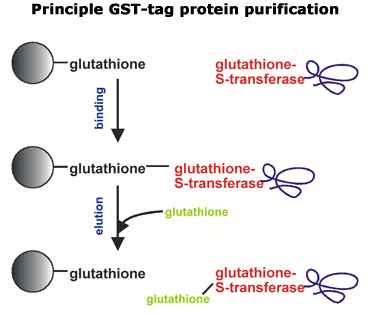
Soluble GST fusion proteins are easily purified using an immobilized glutathione sepharose column. There are several immobilized glutathione chromatography media options available to purify soluble GST fusion proteins from bacterial cell lysates. The protocol described below is adapted from the manufacturer’s recommendation using Glutathione Sepharose 4B poured onto a column and using a peristaltic pump to control flow rates. Protease inhibitors and reducing agents should be added to the buffers as required to minimize proteolysis of the fusion protein. One exception is that serine protease inhibitors must be removed from the glutathione buffer prior to enzymatic removal of the GST moiety, as they will inhibit enzyme activity.

Save a small aliquot from each purification step for SDS-PAGE analysis to monitor fusion protein location throughout the purification. A given column or batch of resin should be used exclusively with a single fusion protein to minimize potential cross-contamination. As an alternative to column purification, a protocol describing batch purifications. Batch purifications are quick and easy, but often the yield and purity of the protein obtained will be somewhat less than that obtained by chromatographic separations. To minimize proteolysis, all protein purification steps should be carried out at 4°C, unless otherwise indicated.

  • Resuspend and pour 20 mL of Glutathione Sepharose 4B resin into a 2.5 × 8 cm column.
  • Thoroughly wash Glutathione Sepharose with 5–10 bed volume PBS at 1.5 mL/min to remove ethanol stock solution.
  • Resuspend pelleted E. coli cells in 15 ml cold Lysis buffer (cells can be fresh or thawed frozen cell pellets).
  • Lyse cells by sonication on ice (~10 times for 10 seconds each with 1-minute rest between bursts to minimize heating of the sample). Save 50 μl of lysate for on-gel analysis and transfer the rest to a 60 ml centrifuge tube.
  • Centrifuge the lysate at 48,000 × g for 20 min at 4 °C.
  • Decant the supernatant into a clean 50 mL centrifuge tube.
  • Resuspend the pellet in 15 ml PBS buffer using a Dounce homogenizer.
  • Run 5-10 μl of the lysate, supernatant, and resuspended pellet on an SDS-PAGE gel to verify that the fusion protein is in the supernatant fraction. If the fusion protein is in the pellet fraction, for tips on improving soluble protein expression for methods of extracting protein from inclusion bodies.
  • Load the soluble fusion protein onto the equilibrated glutathione sepharose column using a flow rate of 0.1 mL/min. Collect fractions and run gels to verify fusion protein binds to column and capacity has not been exceeded. If the fusion protein binds poorly to the resin, for various possible remedies.
  • Wash the column with 5–10 bed volumes of PBS/EDTA/PMSF using a flow rate of 1.5 mL/min.
  • Wash the column with 10-bed volumes of PBS/EDTA using a flow rate of 1.5 mL/min.
  • Elute the fusion protein with glutathione buffer using a flow rate of 0.3 mL/min. Fractions can be monitored using A280 and SDS-PAGE analysis. Pool fractions containing the GST fusion protein. The protein can be stored at 4°C and should be ~90% pure at this stage. If problems are found eluting the fusion protein. If high levels of contamination are present, for troubleshooting tips.

Enzymatic cleavage to remove the GST affinity tag

Depending on the vector chosen, the GST affinity tag can be removed with thrombin, factor Xa, or PreScission protease, either in solution or while still bound to the column matrix. Solution cleavage offers the advantage of greater control over optimizing cleavage conditions, such as temperature, enzyme-to-substrate ratio, incubation duration, and buffer conditions. An advantage of column cleavage is the high level of purity obtained, but this comes at the expense of generally low yield due to less efficient cleavage by proteases and less control of digestion conditions.

Digestion can be performed in the glutathione buffer used to elute the protein from the affinity matrix as long as there are no serine protease inhibitors in this buffer. After incubation, the enzyme can be inhibited using a variety of protease inhibitors or removed using a HiTrap Benzamidine column. Separation of the target protein and the GST moiety can be achieved by further chromatography on the glutathione Sepharose column (after dialysis in PBS buffer) to remove GST and any uncleaved fusion protein. For information on the digestion of GST fusion proteins while bound to column matrix (use with PreScission Protease is recommended).

  • Add the appropriate amount of thrombin or factor Xa to the affinity purified fusion protein and incubate at 37°C (thrombin) or 25°C (factor Xa) for the desired time.
  • Inactivate the enzyme by adding 0.3 mM PMSF (final concentration) to the sample. To ensure complete inhibition, incubate the sample for 15 min at 37°C for thrombin or 30 min at 25°C for factor Xa.
  • Dialyze the sample against PBS/EDTA/PMSF twice using 2 L per dialysis for a minimum of 4 h for each dialysis.
  • Centrifuge the dialyzed sample for 20 min at 4,000 × g to remove any precipitated material that may have formed during the digestion or dialysis steps. At this point, the sample can be reapplied to the glutathione Sepharose column to remove the remaining GST and any undigested fusion proteins.

.

Cusabio Apoptosis Recombinants

Abstract

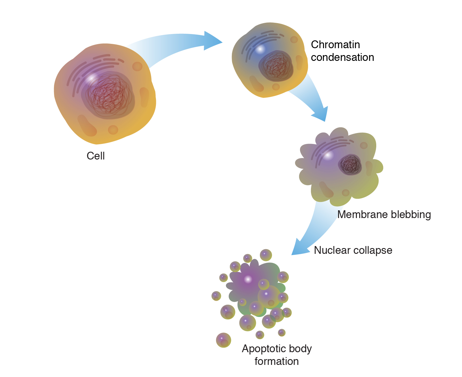
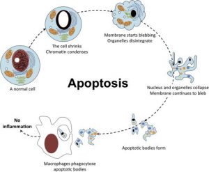
The process of programmed cell death, or apoptosis, is generally characterized by distinct morphological features and energy-dependent biochemical mechanisms. Apoptosis Recombinants is considered a vital component of several processes including normal cell turnover, proper development and function of the immune system, hormone-dependent atrophy, embryonic development, and chemical-induced cell death. Inappropriate apoptosis (either too little or too much) is a factor in many human conditions, including neurodegenerative diseases, ischemic damage, autoimmune disorders, and many types of cancer. The ability to modulate the life or death of a cell is recognized for its immense therapeutic potential.

Therefore, research continues to focus on the elucidation and analysis of the cell cycle machinery and signalling pathways that control cell cycle arrest and apoptosis. To that end, the field of apoptosis research has advanced at an alarmingly fast rate. Although many of the key apoptotic proteins have been identified, the molecular mechanisms of action or inaction of these proteins remain to be elucidated. The objective of this review is to provide an overview of current knowledge about the apoptosis process, including morphology, biochemistry, the role of apoptosis in health and disease, detection methods, as well as a discussion of possible ways apoptotic alternatives.

Keywords: Apoptosis, programmed cell death, intrinsic/extrinsic pathway, granzyme A/B, perforin, autophagy

Distinguish apoptosis from necrosis

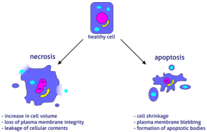
The alternative to apoptotic cell death is necrosis, which is considered a toxic process in which the cell is a passive victim and follows an energy-independent mode of death. But since necrosis refers to the degradative processes that occur after cell death, it is considered by some to be an inappropriate term to describe a mechanism of cell death. Therefore, oncosis is used to describe a process that leads to necrosis with karyolysis and cell swelling, while apoptosis leads to cell death with cell shrinkage, pyknosis, and karyorrhexis.

Therefore, the terms “oncotic cell death” and “oncotic necrosis” have been proposed as alternatives to describe cell death that is accompanied by cellular inflammation, but these terms are not widely used at this time (Majno and Joris, 1995; Levin et al., 1999). Although the mechanisms and morphologies of apoptosis and necrosis differ, these two processes overlap. Evidence indicates that necrosis and apoptosis represent morphological expressions of a shared biochemical network described as the “apoptosis-necrosis continuum” (Zeiss, 2003).

For example, two factors that will convert an ongoing apoptotic process into a necrotic process include a decrease in the availability of caspases and intracellular ATP (Leist et al., 1997; Denecker et al., 2001). Whether a cell dies by necrosis or apoptosis depends in part on the nature of the cell death signal, the type of tissue, the stage of tissue development, and the physiological milieu (Fiers et al., 1999; Zeiss, 2003).

Using conventional histology, it is not always easy to distinguish apoptosis from necrosis, and they can occur simultaneously depending on factors such as the intensity and duration of the stimulus, the degree of ATP depletion, and the availability of caspases (Zeiss, 2003). Necrosis is a passive and uncontrolled process that usually affects large fields of cells, while apoptosis is controlled and energy-dependent and can affect individual cells or groups of cells. Necrotic cell injury is mediated by two main mechanisms; interference with the cell’s energy supply and direct damage to cell membranes.

Physiological apoptosis

The role of apoptosis in normal physiology is as significant as that of its counterpart, mitosis. It demonstrates a complementary but opposite role to mitosis and cell proliferation in the regulation of various cell populations. It is estimated that to maintain homeostasis in the adult human body, about 10 billion cells are produced each day just to balance those that die by apoptosis (Renehan et al., 2001). And that number can increase significantly when apoptosis increases during normal development and ageing or during disease.

Apoptosis is critically important during various developmental processes. For example, both the nervous system and the immune system arise through the overproduction of cells. This initial overproduction is followed by the death of those cells that fail to establish functional synaptic connections or productive antigen specificities, respectively (Nijhawan et al., 2000; Opferman and Korsmeyer, 2003).

Apoptosis is also necessary to rid the body of cells invaded by pathogens and is a vital component of wound healing, as it is involved in the removal of inflammatory cells and the evolution of granulation tissue into scar tissue (Greenhalgh, 1998). . Dysregulation of apoptosis during wound healing can lead to pathological forms of scarring, such as excessive scarring and fibrosis. Apoptosis is also required to eliminate activated or autoaggressive immune cells either during maturation in central lymphoid organs (bone marrow and thymus) or in peripheral tissues (Osborne, 1996).

In addition, apoptosis is critical for remodellings in the adult, such as follicular atresia of the postovulatory follicle and postweaning mammary gland involution, to name a few examples (Tilly, 1991; Lund et al., 1996). Also, as organisms age, some cells begin to deteriorate at a faster rate and are eliminated by apoptosis. One theory is that oxidative stress plays a major role in the pathophysiology of age-induced apoptosis through accumulated free radical damage to mitochondrial DNA (Harman, 1992; Ozawa, 1995). It is clear that apoptosis has to be tightly regulated, as too little or too much cell death can lead to pathologies, including developmental defects, autoimmune diseases, neurodegeneration or cancer.

Apoptosis inhibition

There are many pathological conditions that exhibit excessive apoptosis (neurodegenerative diseases, AIDS, ischemia, etc.) and can therefore benefit from artificial inhibition of apoptosis. As our understanding of the field evolves, the identification and exploitation of new targets remain a considerable focus of attention (Nicholson, 2000). A shortlist of possible methods of anti-apoptotic therapy includes stimulation of the IAP (inhibitor of apoptosis) protein family, caspase inhibition, PARP (poly [ADP-ribose] polymerase) inhibition, stimulation of the PKB/Akt (protein kinase B) pathway and inhibition of Bcl-2 proteins.

The IAP family of proteins is perhaps the most important regulator of apoptosis due to the fact that it regulates both the intrinsic and extrinsic pathways (Deveraux and Reed, 1999). Eight human IAP proteins have now been identified, although XIAP (X-linked mammalian apoptosis inhibitory protein) and survivin remain the best-known members (Silke et al., 2002; Colnaghi et al., 2006). Until now, members of the IAP family have been investigated as therapeutic targets for the treatment of stroke, spinal cord injury, multiple sclerosis, and cancer.

The synthetic non-specific caspase inhibitor z-VAD-fmk was shown to reduce the severity of myocardial reperfusion injury in rat and mouse models of myocardial infarction (Mocanu et al., 2000). Specific inhibitors of caspase activity may also be of benefit. ICE (interleukin-1 beta converting enzyme), also called caspase I, is a cysteine ​​protease that appears to mediate intracellular protein degradation during apoptosis (Livingston, 1997). ACE inhibitors have been developed to treat rheumatoid arthritis and other inflammatory conditions by reducing interleukin 1β (Le and Abbenante, 2005).

Due to the dual role of PARP-1 in both DNA repair and apoptosis, pharmacological use of PARP-1 inhibitors may attenuate ischemic and inflammatory cell and organ injury or may increase the cytotoxicity of antitumor agents ( Graziani and Szabo, 2005). Recent research with PARP-1 knockout mice indicates that the use of PARP-1 inhibitors may be an effective therapy for injury associated with myocardial ischemia and reperfusion injury (Zhou et al., 2006). Infusion of insulin-like growth factor 1 (IGF-1), which stimulates PKB/Akt signalling and promotes cell survival, was shown to be beneficial in animal models of myocardial ischemia (Fujio et al., 2000).

Other studies with transgenic models of cardiac ischemia and global cerebral ischemia indicate that inhibition of Bax expression and/or function can prevent cytochrome c release from mitochondria, inhibit mitochondrial membrane potential lowering, and protect cells against apoptosis (Hochhauser et al., 2003; Hetz et al., 2005). The potential therapeutic modalities mentioned here represent just a few of the past and current research efforts in this field. As the molecular and biochemical complexities of apoptosis are elucidated, new therapeutic strategies will continue to evolve.

Conclusions

Apoptosis is considered a carefully regulated energy-dependent process, characterized by specific morphological and biochemical features in which caspase activation plays a central role. Although many of the key apoptotic proteins that are activated or deactivated in apoptotic pathways have been identified, the molecular mechanisms of action or activation of these proteins are not fully understood and are the subject of continuing research.

The importance of understanding the mechanical machinery of apoptosis is vital because programmed cell death is a component of both health and disease, being initiated by various physiological and pathological stimuli. Furthermore, the widespread involvement of apoptosis in the pathophysiology of disease lends itself to therapeutic intervention at many different checkpoints. Understanding the mechanisms of apoptosis and other variants of programmed cell death at the molecular level provides deeper insights into various disease processes and thus may influence therapeutic strategy.


Animal serum, sera. Siero di vitello fetale e altre specie
 Serum


• Efficienza grazie a lotti preselezionati per ibridomi, cellule ES,
cellule dendritiche ...
• Prenotazione e conservazione del lotto fino a 12 mesi.
• Unico produttore francese - distributore (approvazione veterinaria N ° FR 91 692 200).
• Diverse specie e trattamenti disponibili.
L'efficacia del siero fetale di vitello è essenziale per la tua coltura cellulare.
I nostri test microbiologici, biochimici e di crescita cellulare consentono a Eurobio-Ingen di fornirti i lotti migliori.
SIERO DI SPECIE BOVINA - IL BOVINO SARÀ

Numeri catalogo

CBVBOV00-01 * Siero Bovino 500 ml
CBVVNN00-0U * Siero di vitello neonato 100 ml
CBVVNN00-01 * Siero di vitello neonato 500 ml
CBVCLF00-0U * Siero di vitello 100 ml
CBVCLF00-01 * Siero di vitello 500 ml
CVFSVF06-0U * Siero fetale di vitello decomposto 100 ml
CVFSVF06-01 * Siero fetale di vitello decomposto 500 ml
CVFSVF00-0U * Siero fetale di vitello Eutroph® 100 ml
CVFSVF00-01 * Siero fetale di vitello Eutroph® 500 ml

Regenerative therapy: cell dust to absorb digestive fistulas

Stem cells open up new medical perspectives, but their use is not without risk, in particular because these cells retain their ability to differentiate and divide, which can generate undesirable lines and possibly tumors. In this context, the use of vesicles produced by these cells offers an interesting alternative. Released in the extracellular medium, these vesicles, long regarded as uninteresting “cell dust”, are in fact endowed with therapeutic properties similar to those of mother cells, without presenting the same drawbacks. They have already been used successfully to treat kidney, heart and liver damage. A French research team has just demonstrated for the first time, in pigs, their interest in the treatment of digestive fistulas. These pathologies, consecutive for example to a surgical operation or to certain autoimmune pathologies like Crohn’s disease, create an abnormal communication of the organs of the digestive tract between them or with the skin. The treatments currently available are struggling to absorb them satisfactorily. Gabriel Rhami, of the AP-HP department of gastroenterology and endoscopy, and his colleagues incorporated extracellular vesicles from mesenchymal stem cells into a biocompatible polymer. Injected locally into the digestive fistula at low temperature (4 ° C), this mixture gels with body heat and fills the lesion. The results are spectacular, with a 100% success rate in animals. Researchers are now planning to test the effectiveness of this treatment in the management of perineal fistulas and to perform the first trials in humans.

A.K.A Silva et al. (2018) ACS Nano, 12 (10), 9800-14

8 nuovi strumenti per l’imaging di cellule vive fluorescenti: scopri le sonde SPY

Con il grande successo delle sonde SIR per un’etichettatura efficiente del citoscheletro cellulare, del DNA e del lisosoma, Spirochrome ha appena lanciato una serie di 8 nuovi strumenti fluorescenti a cellule vive: le sonde SPYTM.

Queste 8 nuove sonde SPYTM, recentemente sviluppate da Spirochrome, sono nuovi strumenti di colore (verde, arancione, rosso e rosso intenso – fig.1) che consentono un’etichettatura efficiente di DNA (nucleo), actina e citoscheletro su cellule viventi e fisse. Mantenendo tutte le preziose proprietà delle sonde SIR (non citotossiche, permeabili alle cellule, altamente selettive, compatibili STED / SIM (fig. 2)), queste 8 sonde consentono un’etichettatura forte e accurata dei rispettivi target senza la necessità di manipolazione genetica o sovraespressione di proteine ​​fluorescenti. Grazie alle nuove sonde SPYTM, avrai nuove possibilità di etichettatura per migliorare la qualità dei tuoi dati.

Guarda le immagini dei risultati ottenuti con le sonde SPY ™.
Scopri ulteriori dettagli su queste 8 nuove sonde SPYTM di seguito.

Etichettatura del DNA
SPY505-DNA

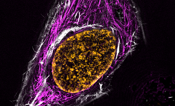
Sinistra: cella HeLa colorata con 1x SPY505-DNA / Destra: cella HeLa colorata con 1x SPY505-DNA (verde) e 1x SPY555-actina (magenta)
Questa colorazione nucleare verde vivo e non tossica per cellule vive consente una rapida etichettatura del DNA in cellule vive e fisse con alta specificità e fondo molto basso (fig. 3). Gli spettri di assorbanza ed emissione sono simili alla fluoresceina o Alexa488TM: (EX. Max: 512 nm; Em. Max: 531nm).

SPY505-DNA può essere ripreso con set di filtri standard fluoresceina (FITC) o YFP e può essere utilizzato per imaging a campo ampio, confocale, SIM o STED in cellule e tessuti viventi o fissi. È compatibile con l’imaging multicolore quando combinato con le altre sonde SPYTM (SPY555, SPY595, SPY650, SPY700), la serie di sonde SiR e RFP o mcherry (fig. 3).

SPY555-DNA

Si tratta di una colorazione nucleare a cellule vive brillante e non tossica che consente una rapida etichettatura del DNA in cellule vive e fisse con alta specificità e fondo molto basso (fig. 4). Gli spettri di assorbanza ed emissione sono simili alla tetrametil-rodamina (TMR): (ad es. Max: 555 nm; Em. Max: 580 nm).

SPY555-DNA può essere ripreso con set di filtri TMR o Cy3 standard e può essere utilizzato per l’imaging a campo ampio, confocale, SIM o STED in cellule e tessuti viventi o fissi. Consente l’imaging multicolore con una qualsiasi delle altre sonde SPYTM (SPY505, SPY595, SPY650, SPY700), la serie di sonde SiR e il GFP.

SPY595-DNA:

Sinistra: cellule HeLa colorate con 1x SPY595-DNA / Destra: cellule HeLa vive colorate con SPY555-actina (magenta), SPY595-DNA (arancione) e SPY650-tubulina (blu)
Questa colorazione nucleare a cellule vive rosso vivo e non tossico consente una rapida etichettatura del DNA in cellule vive e fisse con alta specificità e fondo molto basso (fig. 5).

Può essere ripreso con set di filtri Alexa594TM o Texas Red standard (ad es. Max: 599 nm; Em. Max: 615 nm) e può essere utilizzato per imaging a campo largo, confocale, SIM o STED in cellule e tessuti viventi o fissi. SPY595-DNA consente l’imaging multicolore con le altre sonde SPYTM (SPY505, SPY555, SPY650, SPY700), la serie di sonde SiR e il GFP (fig. 5)

SPY650-DNA

Cellule LeftBJ colorate con 1x SPY650-DNA / Right: cellule HeLa che esprimono EGFP-tubulina (verde) colorate con 1x SPY650-DNA (rosso)
Si tratta di una colorazione nucleare a cellule viventi rosso vivo e non tossica basata sulla serie di coloranti SPYTM che consente una rapida etichettatura del DNA in cellule vive e fisse con elevata specificità e fondo molto basso (fig. 6). L’emissione in rosso riduce al minimo la fototossicità e l’autofluorescenza del campione: (ad es. Max: 652 nm; Em. Max: 674 nm). Il principale miglioramento di questo SPY650-DNA è che a concentrazioni uguali, colora i nuclei cellulari molto meglio del SiR-DNA (10 volte più luminoso senza aumento della tossicità) (fig.7).

Può essere ripreso con set di filtri Cy5 standard e può essere utilizzato per l’imaging a campo ampio, confocale, SIM o STED in cellule e tessuti viventi o fissi. SPY650-DNA consente l’imaging multicolore con le altre sonde SPYTM (SPY505, SPY555, SPY595, SPY700), GFP o m-cherry (fig.6).

Si tratta di una colorazione nucleare a cellule viventi rosso vivo non tossica, che consente una rapida etichettatura del DNA in cellule vive e fisse con elevata specificità e fondo molto basso (fig. 8). Sembra che a uguale concentrazione colora i nuclei cellulari molto meglio del SiR700-DNA (3-6 volte più luminoso senza aumento della tossicità).

La sua assorbanza ed emissione sono simili a Cy5.5: (ad es. Max: 696 nm; Em. Max: 718 nm). SPY700-DNA può essere riprodotto con set di filtri Cy5 ma è meglio usare un filtro di emissione LP a 700 nm e può essere utilizzato per l’imaging a campo ampio, confocale, SIM o STED in cellule e tessuti viventi o fissi.

Etichettatura della tubulina
SPY555-tubulina

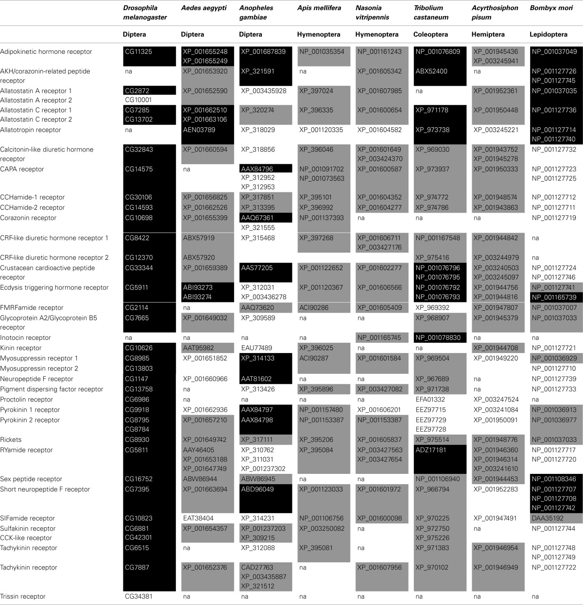
More than two decades of research on insect neuropeptide GPCRs: an overview.

This evaluation focuses on the state of the artwork on neuropeptide receptors in bugs. Most of these receptors are G protein-coupled receptors (GPCRs) and are concerned within the regulation of just about all physiological processes throughout an insect’s life.

More than 20 years in the past a milestone in invertebrate endocrinology was achieved with the characterization of the primary insect neuropeptide receptor, i.e., the Drosophila tachykinin-like receptor. However, it took till the discharge of the Drosophila genome in 2000 that research on neuropeptide receptors boosted.

In the final decade a plethora of genomic info of different insect species additionally grew to become accessible, resulting in a greater perception within the features and evolution of the neuropeptide signaling programs and their intracellular pathways. It grew to become clear that some of these programs are conserved amongst all insect species, indicating that they fulfill essential roles of their physiological processes.

Meanwhile, different signaling programs appear to be misplaced in a number of insect orders or species, suggesting that their actions had been superfluous in these bugs, or that different neuropeptides have taken over their features.

It is putting that the deorphanization of neuropeptide GPCRs will get a lot consideration, however the subsequent unraveling of the intracellular pathways they elicit, or their physiological features are sometimes hardly examined.

Especially in bugs apart from Drosophila this info is scarce if not absent. And though nice progress made in characterizing neuropeptide signaling programs, even in Drosophila a number of predicted neuropeptide receptors stay orphan, awaiting for his or her endogenous ligand to be decided. The current evaluation offers a précis of the insect neuropeptide receptor research of the final two decades.

But it must be emphasised that the work executed up to now is simply the tip of the iceberg and our complete understanding of these necessary signaling programs will nonetheless improve considerably within the coming years.

More than two decades of research on insect neuropeptide GPCRs: an overview.

Roles of glial cells in neural circuit formation: insights from research in bugs.

Investigators through the years have famous many putting similarities within the structural group and performance of neural circuits in increased invertebrates and vertebrates. In newer years, the invention of similarities within the mobile and molecular mechanisms that information growth of these circuits has pushed a revolution in our understanding of neural growth.

Cellular mechanisms found to underlie axon pathfinding in grasshoppers have guided productive research in mammals. Genes found to play key roles within the patterning of the fruitfly’s central nervous system have subsequently been discovered to play key roles in mice.

The range of invertebrate species gives to investigators quite a few alternatives to conduct experiments which are tougher or inconceivable to do in vertebrate species, however which are more likely to shed gentle on mechanisms at play in growing vertebrate nervous programs.

These experiments elucidate the broad suite of mobile and molecular interactions which have the potential to affect neural circuit formation throughout species.

Here we focus on what is thought about roles for glial cells in some of the necessary steps in neural circuit formation in experimentally advantageous insect species. These steps embody axon pathfinding and matching to targets, dendritic patterning, and the sculpting of synaptic neuropils.

A constant theme is that glial cells work together with neurons in two-way, reciprocal interactions. We emphasize the impression of research carried out in bugs and discover how insect nervous programs would possibly greatest be exploited subsequent as scientists search to grasp in but deeper element the complete repertory of features of glia in growth.

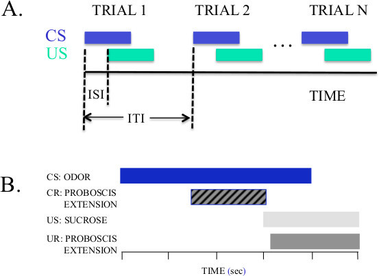
A proboscis extension response protocol for investigating behavioral plasticity in insects: application to basic, biomedical, and agricultural research.

Insects modify their responses to stimuli by way of expertise of associating these stimuli with occasions necessary for survival (e.g., meals, mates, threats). There are a number of behavioral mechanisms by way of which an insect learns salient associations and relates them to these occasions. It is necessary to perceive this behavioral plasticity for applications aimed towards aiding bugs which can be useful for agriculture.

This understanding may also be used for discovering options to biomedical and agricultural issues created by bugs that act as illness vectors and pests.

The Proboscis Extension Response (PER) conditioning protocol was developed for honey bees (Apis mellifera) over 50 years in the past to examine how they understand and find out about floral odors, which sign the nectar and pollen sources a colony wants for survival.

The PER process gives a strong and easy-to-employ framework for learning a number of totally different ecologically related mechanisms of behavioral plasticity. It is definitely adaptable for use with a number of different insect species and different behavioral reflexes. These protocols might be readily employed in conjunction with numerous means for monitoring neural exercise in the CNS by way of electrophysiology or bioimaging, or for manipulating focused neuromodulatory pathways.

It is a sturdy assay for quickly detecting sub-lethal results on conduct attributable to environmental stressors, toxins or pesticides. We present how the PER protocol is simple to implement utilizing two procedures. One is appropriate as a laboratory train for college students or for fast assays of the impact of an experimental remedy.

The different gives extra thorough management of variables, which is necessary for research of behavioral conditioning.

We present how a number of measures for the behavioral response starting from binary sure/no to extra steady variable like latency and length of proboscis extension can be utilized to take a look at hypotheses. And, we talk about some pitfalls that researchers generally encounter after they use the process for the primary time.

A proboscis extension response protocol for investigating behavioral plasticity in insects: application to basic, biomedical, and agricultural research.

Insect speciation guidelines: unifying ideas in speciation analysis.

The examine of speciation is anxious with understanding the connection between causes of divergent evolution and the origin and upkeep of boundaries to gene alternate between incipient species.

Although the sector has traditionally centered both on examples of latest divergence and its causes or on the genetic foundation of reproductive isolation between already divergent species, present efforts search to unify these two approaches.

Here we combine these views by way of a dialogue of latest progress in a number of insect speciation mannequin techniques. We deal with the evolution of speciation phenotypes in every system (i.e., these phenotypes causally concerned in decreasing gene stream between incipient species), drawing an express connection between trigger and impact (course of and sample). We emphasize rising insights into the genomic structure of speciation in addition to well timed areas for future analysis.

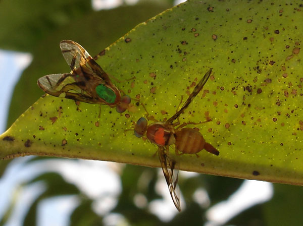
Genetics and biology of Anastrepha fraterculus: research supporting the use of the sterile insect technique (SIT) to control this pest in Argentina.

Two species of true fruit flies (taxonomic household Tephritidae) are thought of pests of fruit and vegetable manufacturing in Argentina: the cosmopolitan Mediterranean fruit fly (Ceratitis capitata Wiedemann) and the new world South American fruit fly (Anastrepha fraterculus Wiedemann).

The distribution of these two species in Argentina overlaps north of the capital, Buenos Aires. Regarding the control of these two pests, the various geographical fruit producing areas in Argentina are in completely different fly control conditions.

One half is underneath a programme utilizing the sterile insect technique (SIT) for the eradication of C.

capitata, as a result of A. fraterculus shouldn’t be current in this space. The software of the SIT to control C. capitata north of the current line with the chance of A.

fraterculus occupying the area of interest left vacant by C. capitata turns into a trigger of a lot concern. Only preliminary steps have been taken to examine the genetics and biology of A.

fraterculus. Consequently, solely fragmentary data has been recorded in the literature relating to the use of SIT to control this species. For these causes, the research to develop a SIT protocol to control A. fraterculus is significantly wanted. In latest years, research teams have been constructing a community in Argentina in order to deal with explicit points of the growth of the SIT for Anastrepha fraterculus.

The issues being addressed by these teams embody enchancment of synthetic diets, facilitation of insect mass rearing, radiation doses and circumstances for insect sterilisation, primary data supporting the growth of males-only strains, discount of male maturation time to facilitate releases, identification and isolation of chemical communication indicators, and a superb deal of inhabitants genetic research.

This paper is the product of a concerted effort to collect all this data scattered in quite a few and typically hard-to-access studies and papers and summarize their primary conclusions in a single publication.

Genetics and biology of Anastrepha fraterculus: research supporting the use of the sterile insect technique (SIT) to control this pest in Argentina.

[Research progress in chemical communication among insect-resistant genetically modified plants, insect pests and natural enemies].

Semiochemicals launched by crops or bugs play an necessary position in the communication amongst crops, phytophagous bugs and their pure enemies. They thus type a chemical data community which regulates intra- and inter-specific behaviors and sustains the composition and construction of plant and insect communities.

The software of insect-resistant genetically modified (IRGM) crops could have an effect on the chemical communication inside and amongst the tritrophic ranges, and thus trigger disturbances to the biotic neighborhood construction and the stability of the farmland ecosystem.

This has raised issues about the environmental security of IRGM crops and triggered research worldwide.

In the present article we supplied a quick abstract of the chemical communication amongst crops, herbivores and pure enemies; analyzed the potential of IRGM crops to have an effect on the chemical communication between crops and arthropods and the associated mechanisms; and mentioned the present research progress and the future prospects in this area.

We hope that this will promote the research in this area by Chinese scientists and enhance our understanding of the potential results of rising of IRGM crops on the arthropod neighborhood construction.